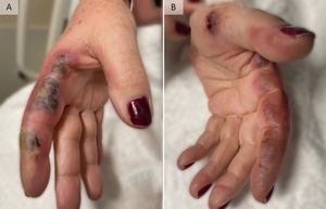
A 66-year-old woman presented to the emergency department with a 1-week history of painful lesions symmetrically distributed on the medial thumbs and lateral index fingers. Physical examination revealed well-demarcated, edematous, violaceous plaques with focal areas of central duskiness (Figure 1A, 1B). The patient had no other skin findings, systemic symptoms, known sick contacts, recent trauma, or new medications. Laboratory testing showed a white cell count of 11,000 per cubic millimeter (73.5% neutrophils), high-sensitivity C-reactive protein of 19.9 mg/L (reference range, ≤3), and erythrocyte sedimentation rate of 42 mm per hour (reference range, ≤20). A punch biopsy of the skin demonstrated a dense neutrophilic infiltrate with negative Gram, Grocott methenamine silver, periodic acid–Schiff, and acid-fast bacterial staining. The patient rapidly improved with initiation of systemic steroids; thus, a diagnosis of neutrophilic dermatosis of the dorsal hands (NDDH), a localized variant of Sweet syndrome, was made.1
Sweet syndrome, also known as acute febrile neutrophilic dermatosis, is considered to be the prototype of the neutrophilic dermatoses, a spectrum of disorders united by a dense, sterile neutrophilic infiltrate on histopathology.2 The diagnosis of classical Sweet syndrome may be made when the acute onset of painful, erythematous nodules or plaques is accompanied by consistent pathology (dense neutrophilic infiltrate without evidence of leukocytoclastic vasculitis) and two of four minor criteria, including excellent response to systemic corticosteroids, presence of fever (>38°C), high inflammatory markers, and association with underlying malignancy, inflammatory disease, pregnancy, or recent illness or vaccination.3 Numerous clinical variants of Sweet syndrome have been described, including pustular Sweet syndrome, necrotizing Sweet syndrome, and NDDH.2 NDDH is a localized variant of Sweet syndrome where skin lesions are usually limited to the dorsa of the hands and often located in the area between the thumb and index finger.1 The skin lesions are classically tender, erythematous to violaceous plaques that become bullous, ulcerative, or pustular.1 Prior to 2006, the relationship between NDDH and diagnostic entities with similar histopathology and clinical characteristics (i.e. pustular vasculitis of the hands, atypical pyoderma gangrenosum) was unclear.4 Now, NDDH is viewed as identical to atypical pyoderma gangrenosum, also called vesiculobullous pyoderma gangrenosum, when that condition presents on the hands.4
The differential diagnosis for NDDH is broad and includes infectious, neoplastic, and autoimmune etiologies.3 Prompt recognition of NDDH is important as central necrosis and ulceration can be confused for a necrotizing infection, which may lead to unnecessary digit resections.5 As seen in other neutrophilic dermatoses, Sweet syndrome can demonstrate pathergy in up to 25% of cases.2 Therefore, patients with NDDH who undergo debridement or resection may experience expansion after surgery.5 Various corticosteroid treatment strategies (including both systemic and topical steroids) for NDDH have been trialed in individual reports, with clinical improvement observed in all cases and resolution of lesions seen usually within a few weeks.1
Approximately 78% of patients with NDDH present with bilateral disease, with only 4% of patients having palmar involvement.1 As in classical Sweet syndrome, there is a significant association between NDDH and underlying (sometimes occult) systemic diseases, including hematologic disorders in approximately 14% of patients, solid tumors in approximately 11% of patients, rheumatologic diseases in approximately 10% of patients, and inflammatory bowel disease in approximately 3% of patients.1 This patient had a history of early-stage renal cell carcinoma with nephrectomy occurring five years prior to onset of symptoms; however, repeat CT imaging of the abdomen and pelvis after the diagnosis of NDDH did not show recurrence of disease.
Author Contributions
All authors have reviewed the final manuscript prior to submission. All the authors have contributed significantly to the manuscript, per the International Committee of Medical Journal Editors criteria of authorship.
-
Substantial contributions to the conception or design of the work; or the acquisition, analysis, or interpretation of data for the work; AND
-
Drafting the work or revising it critically for important intellectual content; AND
-
Final approval of the version to be published; AND
-
Agreement to be accountable for all aspects of the work in ensuring that questions related to the accuracy or integrity of any part of the work are appropriately investigated and resolved.
Disclosures/Conflicts of Interest
The authors have no conflicts of interest to disclose.
Corresponding author:
Andrew Sanchez, MD
Yale University Department of Internal Medicine
333 Cedar Street
P.O. Box 208056
New Haven, CT, USA 06510
Ph: 203.688.9503
Email: Andrew.Sanchez@Yale.edu

