A 47-year-old female with history of hypertension, irritable bowel syndrome, fibromyalgia, chronic pain disorder, anxiety, depression, bilateral obstructing renal stones status post recent left ureteral stent and right percutaneous nephrostomy tube placement presented with a 4-week history of rectal bleeding, increasing abdominal pain, and 8-pound weight loss. She was taking buprenorphine-naloxone, oxybutynin, amitriptyline, duloxetine, atenolol, loratadine, depo-provera and cyclobenzaprine. Her CRP was 89 mg/L (ref: 0-10mg/L) and an infectious work-up was negative. A CT abdomen/pelvis done at an outside hospital showed colitis of the sigmoid colon and mild residual right hydroureteronephrosis. Colonoscopy revealed moderate inflammation from the rectum to the ascending colon. Pathology showed mild to moderate colitis with areas of erosion, and cytomegalovirus testing was negative. She was diagnosed with ulcerative colitis, started on 40mg prednisone daily, and discharged home with close outpatient gastroenterology follow-up.
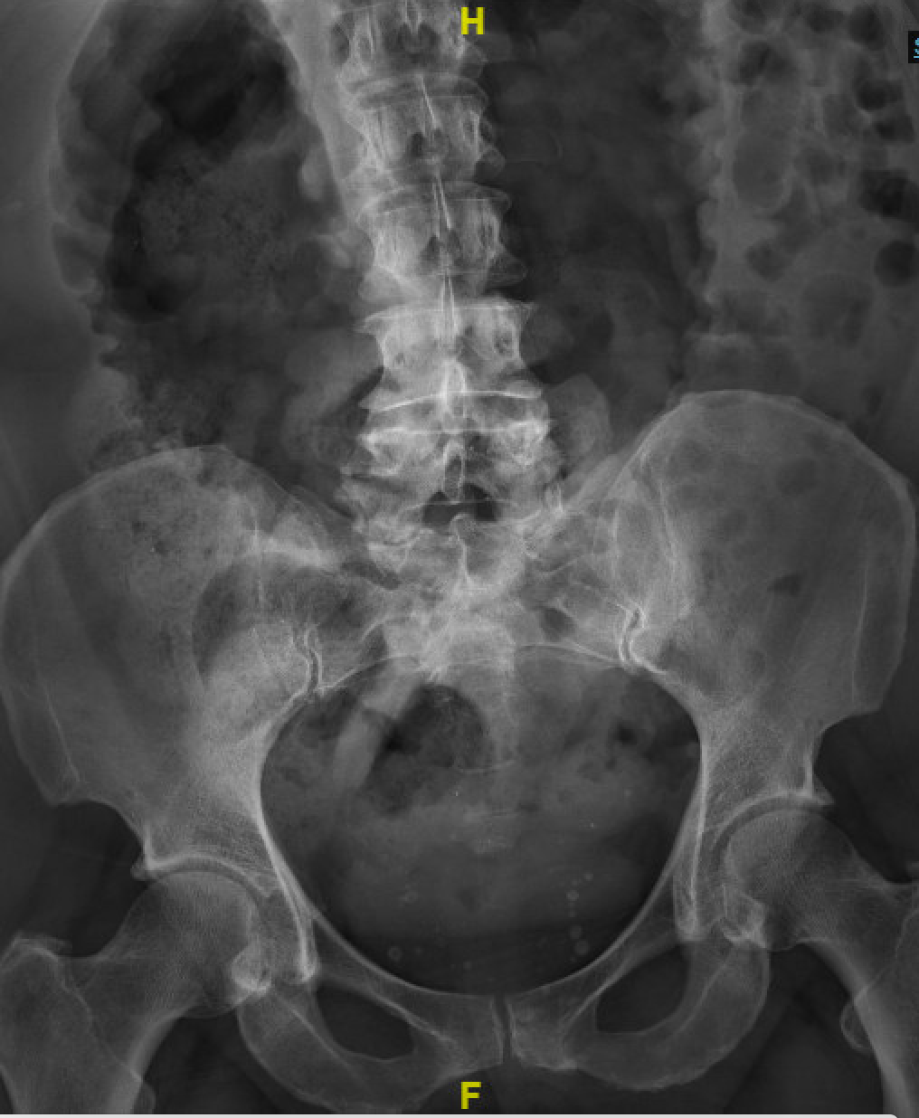
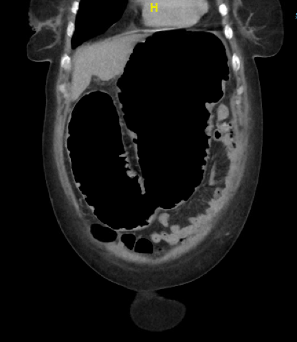
She presented again six days after hospital discharge due to worsening abdominal pain. She also reported alternating between having diarrhea and being constipated. On arrival, she was afebrile and hemodynamically stable. Her laboratory workup revealed white cell count 16,000 (ref: 4200-10000), Hgb 9.0 g/dL (ref: 11.2-14.9g/dl), Cr 1.32 mg/dL (ref: 0.44-1.03mg/dl), albumin 3.5 gm/dl (ref: 3.4-5.1 gm/dl), CRP 172 mg/L (ref: 0-10mg/L), ESR 64 mm/h (ref: 0-20mm/hr). She was treated with IV solumedrol 20mg every 8 hours. On hospital day 2, she reported chest pain for which chest and abdominal X-rays were done, showing a dilated colon measuring up to 7.8 cm at the transverse colon and thumbprinting of the colonic wall (Figure 1). Despite being on steroids, the patient reported having ongoing abdominal pain along with no bowel movement. Given her history of irritable bowel syndrome and chronic pain disorder, it was challenging to ascertain if there was any improvement with the steroids. Flexible sigmoidoscopy was done, and it showed moderate to severe inflammation in the rectum and sigmoid colon. Due to a suboptimal response to steroids, she was given a dose of infliximab on day 5. She initially had symptomatic improvement but developed worsening abdominal distension accompanied by rising CRP to 343 mg/L. A CT abdomen/pelvis on day 9 showed significantly dilated gas-filled loops of large bowel, most pronounced in the transverse colon measuring up to 10.4 cm along with loss of haustral markings and a pseudopolyp concerning for toxic megacolon (Figure 2). Colorectal surgery was consulted, and a total abdominal colectomy was performed with end-ileostomy on the same day with findings of gangrenous, perforated transverse colon and feculent peritonitis.


Toxic megacolon is an acute complication seen in ulcerative colitis, less so in infectious and other types of colitis. It is a fulminant colitis characterized by loss of the colon’s neurogenic tone, leading to severe dilatation and an increased risk of perforation.
In addition to Inflammatory bowel disease, usual causes in adults include:
-
Infectious colitis (such as C. difficile colitis, Shiga-toxin producing E. coli, Shigellosis, Amoebiasis)
-
Radiation-induced colitis
-
Colonic lymphoma
-
Graft-versus-host disease
-
Hypothyroidism
Patients usually present with symptoms such as abdominal bloating, abdominal pain, fever, tachycardia, evidence of dehydration, and signs of hypovolemic or septic shock. In addition to diffuse abdominal tenderness on physical examination, bowel sounds may be hypoactive or absent. CT imaging usually reveals colonic wall thickening, inflammatory pseudopolyps (islands of normal colonic mucosa surrounded by denuded ulcerative mucosa), thumbprinting, and pericolic fat stranding from mucosal edema may be seen.1,2
There are 3 major components in the management of toxic megacolon1:
-
Supportive: Fluid and electrolyte repletion
-
Pharmacologic: Steroids, Biologics (to blunt the response of the inflammatory cytokines and other mediators involved in its pathogenesis), and the avoidance of NSAIDs
-
Surgery
In cases due to inflammatory bowel disease, early identification of features associated with elevated risk of steroid failure is critical to prevent complications such as perforation, fulminant sepsis, and shock. The utility of the Travis or Oxford criteria (based on stool frequency >8 and CRP >45 mg/L, on day 3 of admission) has been questioned due to availability of new treatment modalities.2 According to several studies, mostly retrospective, a combination of the following is associated with a remarkably elevated risk of steroid failure and need for colectomy, in up to 78% of cases3,4:
-
Serum albumin <30g/L on presentation
-
CRP>50 (on day 3 of treatment with Steroids)
-
Fecal calprotectin levels >1922 µg/g
-
UCEIS (Ulcerative colitis endoscopic index of severity) score >7.
With the increasing use of immune checkpoint inhibitors and the potential for increased prevalence of associated cases of autoimmune colitis, these and other criteria for early initiation of step-up therapy must be elucidated. These criteria should be used to identify patients who need the early institution of biologic agents in addition to steroids to help lower colectomy rates and predict patients who may require colectomy to reduce peri-operative complications
AUTHOR CONTRIBUTIONS
All Authors have reviewed the final manuscript prior to submission. All the authors have contributed significantly to the manuscript, per the ICJME criteria of authorship.
-
Substantial contributions to the conception or design of the work; or the acquisition, analysis, or interpretation of data for the work; AND
-
Drafting the work or revising it critically for important intellectual content; AND
-
Final approval of the version to be published; AND
-
Agreement to be accountable for all aspects of the work in ensuring that questions related to the accuracy or integrity of any part of the work are appropriately investigated and resolved
DISCLOSURES/CONFLICTS OF INTEREST
The authors have no conflicts of interest to disclose.
CORRESPONDING AUTHOR
Kwame Dapaah-Afriyie MD
Professor of Medicine, Clinical Educator
Warren Alpert Medical School at Brown University
Division Director
Division of Hospital Medicine
The Miriam Hospital, 164 Summit Avenue, Providence, RI 02906