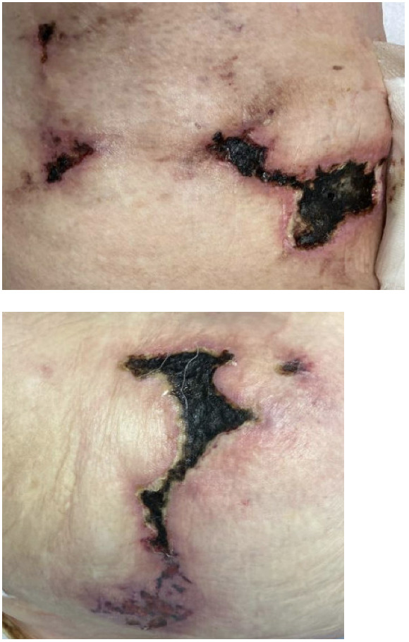

A 78-year-old female with a past medical history significant for heart failure with reduced ejection fraction, atrial fibrillation, aortic stenosis status post mechanical aortic valve replacement treated with warfarin for over ten years presented with painful necrotic skin ulcerations for the past ten months. The patient noted that the lesions began as erythematous migratory blistering skin lesions initially involving the ankles that evolved into necrotic ulcerations, and eventually moved to the shins, thighs, hip, buttock, and abdomen (Figures 1 and 2). Prior to her presentation, the patient was treated with multiple antibiotics along with wound care and debridement in the outpatient setting. These interventions proved to be ineffective, as the skin lesions were enlarging and spreading to new areas, prompting her visit to the hospital for a higher level of care. On arrival, vital signs were stable. A complete blood count and basic metabolic panel were both within normal limits. Prothrombin time (PT) was elevated at 26.2 sec (ref: 12.1-14.8 sec) and the international normalized ratio (INR) was elevated at 3 (ref: 0.8-1.2) in the setting on warfarin usage. Wound cultures were significant for heavy pseudomonas aeruginosa. Blood cultures were negative. Dermatology was consulted for biopsy and treatment recommendations. Biopsies revealed vascular calcium deposition associated with the deep dermis and superficial subcutaneous tissue.


Taken together, these lesions were suggestive of non-uremic calciphylaxis in the setting of warfarin intake. Cardiology was consulted for recommendations regarding warfarin usage for prior mechanical aortic valve replacement in the setting of the newly diagnosed non-uremic calciphylaxis. The patient and her husband were very hesitant to discontinue warfarin as she was on warfarin for over a decade after mechanical aortic valve replacement. Shared decision making was done with the patient and her husband at bedside, and she was subsequently transitioned from warfarin to low-molecular-weight heparin (LMWH). She was also treated with intravenous sodium thiosulfate. The patient was scheduled for follow-up with outpatient dermatology after discharge; however, her hospital course was further complicated by acute hypoxic respiratory failure and she ultimately passed away shortly after.
Calciphylaxis is a rare life-threatening condition most commonly affecting patients with end stage kidney disease on dialysis. It is characterized by calcification and thrombosis of small blood vessels, leading to painful non-healing necrotic skin ulcers, which can be complicated by secondary infection and sepsis, contributing to its high mortality rate.1,2 Unlike uremic calciphylaxis, non-uremic calciphylaxis occur in patients without end-stage renal disease. It is linked to a broad spectrum of underlying conditions such as chronic kidney disease, hyperphosphatemia, hypercalcemia, hyperparathyroidism, and vascular calcification.1–3 Additional associations include coagulation disorders, aluminum toxicity and iron dextran use.1,2 Clinical reports have also suggested potential links with renal transplantation, immunosuppressive therapy, corticosteroid use, and obesity.1,2
Warfarin, a commonly prescribed anticoagulant, has also been associated with the development of calciphylaxis. Warfarin- induced non-uremic calciphylaxis should be considered in patients with normal kidney function presenting with painful necrotic lesions in the setting of chronic warfarin usage. The lesions may initially present as livedo reticularis, reticulate purpura, violaceous plaques, or indurated nodules. Ulcerations often progress to necrotic lesions with black eschar, as observed in our patient. Regardless of etiology, calciphylaxis is associated with high morbidity due to severe pain, chronic non-healing wounds, prolonged hospitalizations, and recurrent superimposed infections. Ulcerated lesions in calciphylaxis carry a reported 1-year mortality rate of 45–80%, with sepsis being the most common cause of death.1,2
Histopathologically, calciphylaxis is marked by microvascular thrombosis, calcification, and fibrointimal hyperplasia of cutaneous arterioles, leading to ischemia and septal panniculitis.1,2,4,5 Vascular calcification is thought to cause endothelial dysfunction and injury; however, the precise pathophysiology, diagnostic criteria, and optimal management of this devastating condition remain incompletely understood.5 Warfarin-induced non-uremic calciphylaxis presents with painful, necrotic skin lesions that are typically located on the lower extremities, particularly below the knees. Patients may experience severe pain, erythematous nodules, and plaques that can progress to ulcerations and gangrene.1 The condition predominantly affects females and is associated with long-term warfarin use, with an average duration of 32 months after initiation of warfarin.2,4
Our patient has many risk factors for the development of non-uremic calciphylaxis, including warfarin use, female sex, and obesity. When considering this patient’s history of chronic warfarin usage in the setting of lower extremity necrotic ulcerative lesions, and the characteristic pathology report, the most likely etiology of these necrotic lesions is warfarin-induced non-uremic calciphylaxis. The pathophysiology of warfarin- induced non-uremic calciphylaxis is driven by the relationship between warfarin and vascular calcification. Warfarin inhibits the vitamin K-dependent carboxylation of matrix Gla protein (MGP), a potent inhibitor of vascular calcification.1,2 This inhibition leads to increased vascular calcification of the tunica media and intima of small vessels.5
Shared decision-making is essential when initiating warfarin therapy in patients with mechanical heart valves, given the serious risks of complications such as warfarin-induced calciphylaxis and the high risk of stroke with anticoagulation nonadherence.6 Treatment of this condition includes discontinuation of warfarin and substitution with heparin or LMWH to halt further progression of the calcifications. Sodium thiosulfate treatment is recommended for its ability to enhance calcium solubility and promote excretion.1–3 Hyperbaric oxygen therapy has also been described to promote wound healing and is associated with better survival when combined with other treatments in patients with warfarin-induced non-uremic calciphylaxis.1–3 Wound care and multimodal pain management with NSAIDs and opioids are also recommended to limit potential infectious complications and mitigate the severe pain associated with calciphylaxis, respectively.
Author Contributions
All authors have reviewed the final manuscript prior to submission. All the authors have contributed significantly to the manuscript, per the International Committee of Medical Journal Editors criteria of authorship.
-
Substantial contributions to the conception or design of the work; or the acquisition, analysis, or interpretation of data for the work; AND
-
Drafting the work or revising it critically for important intellectual content; AND
-
Final approval of the version to be published; AND
-
Agreement to be accountable for all aspects of the work in ensuring that questions related to the accuracy or integrity of any part of the work are appropriately investigated and resolved.
Disclosures/Conflicts of Interest
The authors declare they have no conflicts of interest
Corresponding Author
Farzana Hoque, MD, MRCP, FACP, FRCP
Associate Professor of Medicine, Department of Internal Medicine,
Saint Louis University School of Medicine, Phone: 314-257-8222
Email: farzanahoquemd@gmail.com