

A 72-year-old female with a past medical history significant for collagenous colitis, anemia, anxiety, cervical myelopathy, chronic obstructive pulmonary disease, depression, gastric reflux, hyperlipidemia, hypertension, mitral valve prolapse, obstructive sleep apnea, and Parkinson’s disease presented with new onset rash following antibiotic administration. One week prior to her presentation, the patient was treated for an ear infection with amoxicillin and ciprofloxacin-dexamethasone ear drops. One-hour post-amoxicillin administration, the patient developed an itchy rash on her bilateral hands. Ciprofloxacin-dexamethasone ear drops had been started several hours prior to the reaction. The next day the patient presented to her primary care provider and was switched to doxycycline with a one-time dose of IV Methylprednisolone 60 mg. Despite having previously tolerated amoxicillin without issue, she experienced a rapidly spreading, painful rash associated with fever, chills, nausea, and vomiting for several days that prompted her to visit the hospital.
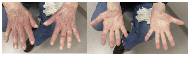
On arrival, the patient was afebrile and hemodynamically stable. Physical exam was notable for painful purplish, erythematous lesions with ruptured bullae located on the dorsum of the hands and wrist with additional small, isolated lesions on bilateral knees (Figure 1). Labs were notable for an elevated white blood cell count of 14,600 per cubic millimeter (reference range 4,000 - 10,700), erythrocyte sedimentation rate of 115 mm per hour (reference range < 30), and C-reactive protein of 28.6 mg/ dl (reference range <0.5). She was evaluated by dermatology and started on prednisone 90 mg due to concern of reactive neutrophilic dermatosis. Skin biopsy of the dorsum of her right hand showed dermal neutrophilic infiltrate with focal leukocytoclastic vasculitis and epidermal necrosis. Immunofluorescent studies of the tissue collected were negative for IgA, IgM, IgG, C3, type IV collagen, and fibrinogen. Anti-neutrophil cytoplasmic antibody vasculitis panel was also negative. Bacterial, fungal, and acid-fast bacilli cultures were collected and found to be negative as well. The patient showed improvement in swelling and pain after two days of treatment and was discharged with Prednisone 80 mg until follow-up with dermatology. Given the timing of symptom presentation shortly after systemic amoxicillin administration, and the localized therapeutic effect of ciprofloxacin-dexamethasone ear drops, it was concluded that amoxicillin administration was likely the inciting factor in the setting of her known history collagenous colitis. This patient’s Naranjo score was found to be 6, suggesting the probability of an adverse drug reaction secondary to amoxicillin administration is probable. The patient was counseled to avoid amoxicillin, and at her two-week follow-up appointment, the patient was found to have substantial improvement in her rash (Figure 2) along with a resolution of pain. Her prednisone was decreased to 60 mg daily, and she was started on clobetasol ointment with additional follow-up in one month. The patient was also referred to the gastrointestinal clinic for management of collagenous colitis, given this condition may have contributed to her presentation.

Reactive neutrophilic dermatosis refers to a group of noninfectious, autoinflammatory skin disorders characterized by the presence of a sterile neutrophilic infiltrate in the skin on histology.1 The prototypical form of neutrophilic dermatosis includes acute febrile neutrophilic dermatosis, also known as Sweet’s syndrome (SS). SS is characterized by the sudden onset of fever, leukocytosis, and tender erythematous skin lesions such as papules, nodules, and plaques. Histologically, SS is marked by a dense infiltrate of mature neutrophils in the upper dermis without evidence of vasculitis.2 Laboratory findings include leukocytosis and elevated inflammatory markers such as erythrocyte sedimentation rate and C- reactive protein. SS most commonly presents in females with the average age of onset between 30 to 60 years age, although several cases have been reported within females in their 70s and 80s.3 SS can be divided into three main subtypes: idiopathic, malignancy associated, and drug induced.4,5 Idiopathic SS is associated with infections, vaccinations, and inflammatory disorders such as inflammatory bowel disease, while malignancy- associated SS is more commonly reported with hematologic malignancies and myeloproliferative or myelodysplastic disorders.5 Drug induced SS is most commonly associated with colony stimulating factors; however, a few cases have reported an association between oral amoxicillin administration and the development of acute febrile neutrophilic dermatosis.6 While other antibiotics have also been implicated, there are no documented cases of SS occurring secondary to the use of ciprofloxacin- dexamethasone ear drops.
Localized variants of SS have also been described, including NDDH. NDDH is characterized by painful erythematous and violaceous papules, plaques, nodules, pustules, and hemorrhagic bullae most commonly located on the dorsal aspect of both hands, with occasional involvement of the wrists, palms, and legs.5 The main difference is that NDDH is a localized, acral variant of SS, with lesions confined to the dorsal hands and a higher frequency of bullae and ulceration, whereas SS is more generalized and often associated with systemic symptoms. While the diagnosis of NDDH requires histological findings that excluded vasculitis, one case published by Mobini et al., described a patient presenting with NDDH and a skin biopsy showing diffuse neutrophilic infiltration of the dermis with leukocytoclastic vasculitic changes.7 Further workup of this patient was negative for vasculitis, and the authors concluded that “the timing of the biopsy during the evolutionary phases of the lesions may result in different findings with regard to presence or absence of vasculitis.7” First line treatment for reactive neutrophilic dermatosis includes systemic steroids; however, treatment of any underlying disorder or malignancy is crucial.5 Other steroid-sparing treatments include potassium iodide, colchicine, and dapsone.5
This case is most consistent with reactive neutrophilic dermatosis, specifically neutrophilic dermatosis of the dorsal hands (NDDH), occurring in the setting of collagenous colitis and likely precipitated by recent amoxicillin exposure. Vasculitis and infectious etiologies were considered in the differential; however, the negative workup rendered these diagnoses less likely. The association between NDDH and systemic inflammatory conditions, including collagenous colitis, highlights the importance of recognizing this entity in similar clinical contexts.
Disclosures/Conflicts of Interest
The author has no conflicts of interest to disclose.
Corresponding Author
Michael Kanan, BSc
Saint Louis University School of Medicine
Saint Louis, MO 63104-1016 USA
Email: michael.kanan@health.slu.edu