
A 65-year-old female with a history of collagenous colitis and a thyroid nodule developed a small patch of erythema on the ventral aspect of the middle finger of her right hand. Over the next two months, the erythema extended to the entire finger, accompanied by edema and warmth, prompting concern for cellulitis. Wound cultures obtained during two surgical debridements subsequently tested positive for Mycobacterium marinum. During this period, her antibiotic regimen included cefuroxime, Bactrim, mupirocin, vancomycin, clarithromycin, and azithromycin. Following the culture results, the antibiotic regimen was adjusted to rifampin, ethambutol, and azithromycin. She denied any recent travel, sick contacts, or insect/animal bites. She enjoyed caring for a saltwater fish tank that she maintained for 30 years. She was the full-time caretaker to her relative and retired healthcare worker.
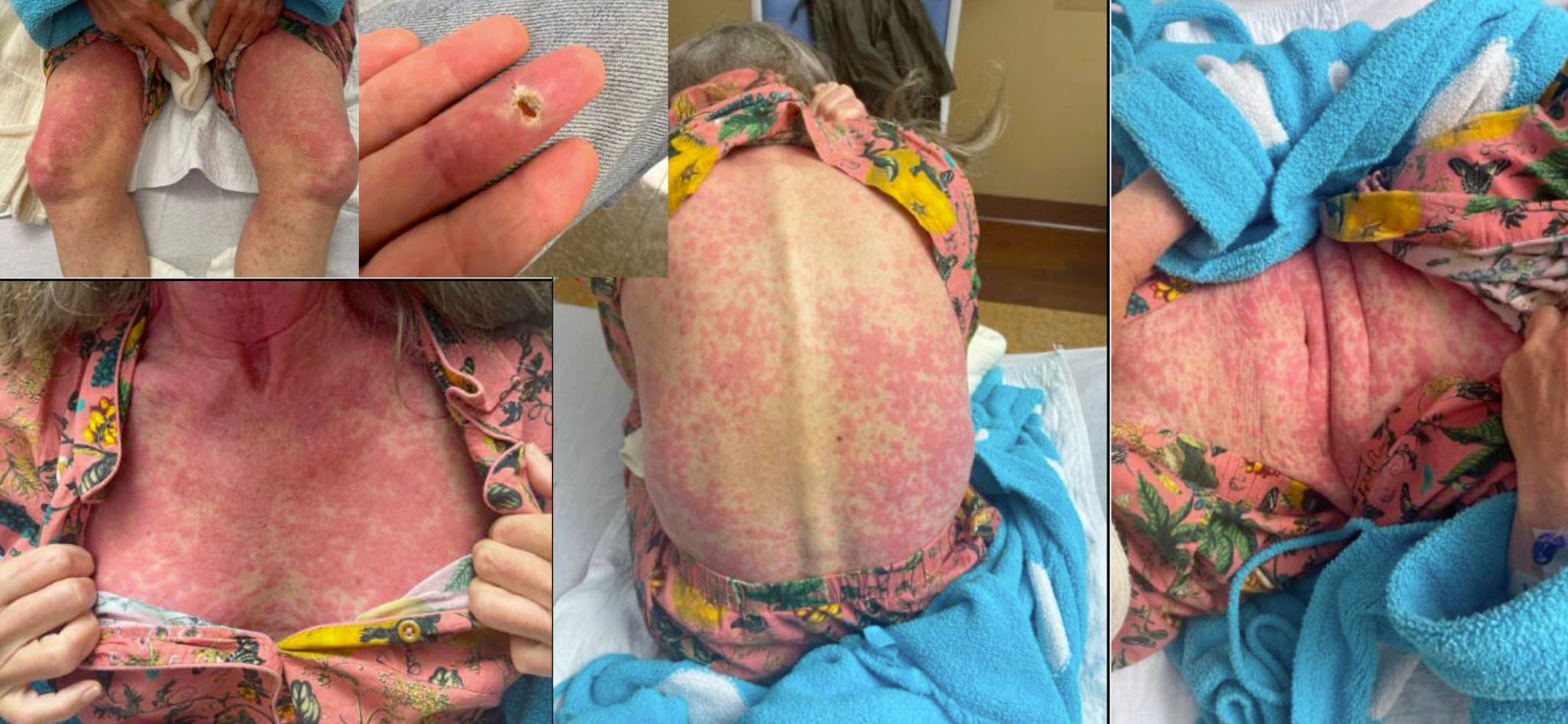
In the following two weeks, she presented herself twice to an outside hospital for ongoing fever and abdominal pain, at which times workup including complete blood count, comprehensive metabolic panel, tick panel, and abdominal computed tomography scan were unrevealing. She then presented for a new non-pruritic rash progressing from the torso to the extremities over the course of a few hours (Figure 1). She also endorsed sore throat, headache, facial and periorbital edema, mild diplopia, and oral ulcers.

On admission to the hospital, she was febrile upto 102.8F and tachycardic. Her exam was notable for swelling and erythema of bilateral eyelids, enlargement of two cervical and axillary lymph nodes, and epigastric tenderness to palpation. Skin exam showed a progressively confluent, erythematous, mildly edematous maculopapular rash extending from the torso to the extremities, sparing the hands and feet and without bullae or desquamation. There was a 1 cm ulcer with central necrosis and surrounding erythema on the ventral aspect of the right middle finger, which was unchanged since the most recent debridement.
Initial labs were notable for leukopenia (3.5 × 10⁹, ref 5-10 × 109/L), hypoalbuminemia (3.2, ref 3.5-5.5 g/dL), and elevated liver enzymes: aspartate transaminase (443, ref <35 U/L), alanine transferase (583, ref <36 U/L), and alkaline phosphatase (149, ref 30-120 U/L). Troponin was <0.1 ng/mL and creatinine was 0.84 mg/dL. Urine analysis and respiratory pathogen panel were negative for infection. A skin biopsy of the rash was collected and showed rare eosinophils admixed within the perivascular lymphocytic infiltrate. Her existing antibiotic regimen was discontinued, and IV methylprednisolone was initiated. For skin discomfort from the rash, she was treated with topical steroids, petroleum jelly, hydroxyzine and diphenhydramine. For oral herpes simplex virus reactivation, she was treated with maalox spit and swish. On hospital day 3, eosinophilia was noted (WBC 3.8 with 4.9% eosinophils, ref 5-10 × 109/L with 1-4% eosinophils). Liver function tests and C-reactive protein levels continued to decline, and there was no evidence of mucocutaneous involvement. She was felt to have drug reaction with eosinophilia and systemic symptoms (DRESS) syndrome, which was triggered by one of the antimicrobial medications she was taking. Considering her improving clinical picture, she was discharged on hospital day 3 with close outpatient infectious disease (ID) follow-up.
The ID team resumed azithromycin 7 weeks later without symptom recurrence. Three weeks after that, ethambutol was resumed. Within hours, she developed a full body erythematous rash, fever, and sore throat and she was readmitted to the hospital. Complete blood count, liver function tests, and inflammatory markers were within normal limits. A high dose prednisone taper was initiated for DRESS syndrome from ethambutol re-exposure. All antimicrobial agents have been held since that time without evidence of infection recurrence. A timeline of the medication exposures and hospitalizations have been described in Figure 2. The necrotic ulcer on her finger healed well, though she endorsed stiffness likely secondary to scar tissue.

DRESS is a rare drug reaction occurring in 1 in 1,000 to 1 in 10,000 drug exposures. It is characterized by a constellation of symptoms including fever, rash, internal organ involvement, and eosinophilia between 2 and 8 weeks after drug exposure.1 It is associated with a 2-10% mortality and permanent end organ damage if treatment is delayed.2 Common offending agents include antiepileptic drugs (particularly carbamazepine), allopurinol, antimicrobial sulfonamides and dapsone, and other antibiotics.3 It most commonly affects middle-aged women and is associated with long-term infectious and autoimmune sequelae, such as herpesviridae reactivation, Type 1 Diabetes, Graves’ disease, and Hashimoto’s thyroiditis.4,5 Cases of severe organ damage requiring liver transplant and temporary renal replacement therapy have been reported.6,7 The pathogenesis of DRESS is not well understood, but hypothesized to be T-cell mediated with a genetic predisposition.8,9 Mycobacterium marinum is a rare cause of cellulitis, acquired by exposure of compromised dermal tissue to contaminated aquatic environments. Though the patient was exposed to multiple potential causative agents, ethambutol was felt to be the likely culprit as her rash recurred with rechallenge. DRESS typically manifests 2 to 8 weeks after initiation of the offending drug. In this case, classic symptoms of rash, fever, transaminitis occurred by day 10 and eosinophilia by day 13. Though all antibiotics were discontinued in treatment of DRESS, M. marinum cellulitis did not progress or recur, possibly facilitated by two earlier surgical debridements.
DRESS is defined by the RegiSCAR criteria with a scoring system of -1, 0, +1 and +2 for each criterion (Table 1). Diagnosis likelihood is described based on points: no case (<2), possible (2-3), probable (4-5), and definite (>5). Definitive treatment includes immediate identification and cessation of the offending agent(s) as well as steroids to reduce inflammation and autoreactivity. In cases such as this one, antimicrobial treatment must also be resumed when appropriate. In the weeks following the primary episode, a nonspecific hypersensitivity reaction to any drug can occur that closely resembles DRESS recurrence.4,9 Reintroduction of the causative agents is generally contraindicated. However, in conditions with limited pharmacological options, patch testing has been explored as a means to help guide treatment decisions.9,10
Anti-tuberculosis medications such as rifampin, ethambutol, isoniazid, and azithromycin are uncommon causes of DRESS, especially compared with agents like allopurinol and carbamazepine. The Adverse Drug Reaction Probability Scale (Naranjo Algorithm) evaluates causality based on exposure timing and clinical progression. In this case, the score was 7, consistent with “probable causality,” whereas a score above 9 would indicate “definite causality.”11 The French Pharmacovigilance System has documented a rise in DRESS cases linked to these agents over the past two decades.⁴ Among 67 reported cases, 10 showed symptom onset earlier than the typical 2–8 week latency period, as observed in this patient. Similarly, case reports present rapid onset DRESS recurrence upon ethambutol rechallenge, quicker than other agents.12,13 Most cases of DRESS associated with ethambutol and rifampin occur in the setting of tuberculosis treatment.4 Though infection with M. marinum is more rare than M. tuberculosis, both treatment paradigms raise interesting considerations in the management of DRESS. In both cases, multiple agents are initiated in tandem, complicating the identification of a causative agent should DRESS arise. Moreover, treatment regimens span months, so agents may be discontinued prematurely in the management of DRESS. Careful consideration must be given in completing the treatment course of the primary infection without reactivating DRESS.
This case is unique in the early onset reaction, atypical infectious source, and rising evidence for the implication of anti-mycobacterial treatment in DRESS. The presumed etiology of her infection was her saltwater fish tank that she cleaned for 30 years without gloves. M. marinum is typically treated with a prolonged course of clarithromycin combined with either ethambutol or rifampin, both of which are known triggers of DRESS. Management relies on identification and discontinuation of the causative agent(s). To our knowledge, this is the first reported case of DRESS in the treatment of M. marinum. Although M. marinum necessitates prolonged antibiotic therapy, this case highlights the potential for life-threatening adverse drug reactions associated with these treatments.
Disclosures/Conflicts of Interest
The authors have no conflicts of interest to disclose.
Corresponding Author
Sonja Kapadia
Warren Alpert Medical School of Brown University
Providence, RI, USA
Email: sonja_kapadia@brown.edu