A 39-year-old man with a prior right anterior cruciate ligament rupture complicated by traumatic arthritis of the right knee presented for an elective right total knee arthroplasty. Per his preoperative airway exam, he was a Mallampati class II. He underwent general endotracheal anesthesia and was successfully intubated on the first attempt with a Macintosh laryngoscopy blade number 4 and a 7.5-mm endotracheal tube (ETT). He was extubated and transferred to the post-anesthesia care unit with stable vital signs on room air.
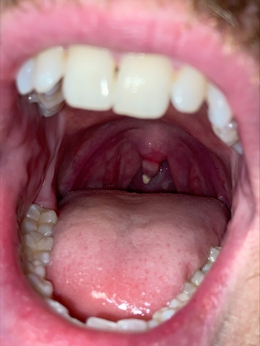
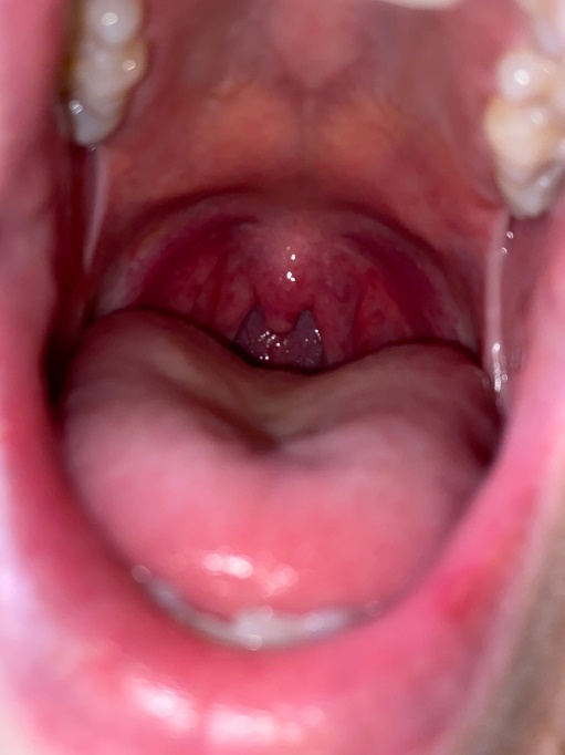
On post-operative day (POD) 4, the patient reported a sore throat, which had persisted since extubation on POD 0. He did not report any dysphagia, odynophagia, hoarseness, or dyspnea. On exam, his uvula was edematous with an ulcerated tip. There was no tonsillar enlargement, exudates, other oral lesions, or cervical lymphadenopathy (Figure 1). He was afebrile with normal vital signs and no leukocytosis. The case was discussed with Otolaryngology, who felt his symptoms were likely due to uvular necrosis related to his recent intubation. Conservative management was recommended, and he was prescribed throat lozenges and phenol spray. His sore throat and uvular edema gradually improved with these measures. By POD 8, the previously seen ulceration on his uvula was no longer appreciated (Figure 2). He was discharged to an acute rehabilitation unit to continue physical therapy.


Sore throat is common in patients who have recently undergone endotracheal intubation. This non-specific pharyngeal irritation is usually rated as mild to moderate and lasts less than two days.1 Uvular ischemia or even necrosis are rare but notable causes of a persistent or severe sore throat following extubation. Uvular ischemia results from impaired blood flow to the uvula often due to compression from midline placement of the ETT, uvular entrapment between the ETT and oral airway, or vigorous suctioning.2 Use of any oropharyngeal instrumentation, whether an artificial airway or suctioning device, is the major risk factor for uvular injury.3 Risk of uvular necrosis does not appear to differ significantly between ETTs and laryngeal mask airways. Male gender is a proposed risk factor as men have more non-fatty tissue in the neck and soft palate. With induction of general anesthesia, this soft tissue becomes flaccid and prone to compression by artificial instrumentation.3,4
Uvular necrosis is diagnosed clinically based on symptoms such as throat pain, dysphagia, or odynophagia in a patient with recent endotracheal intubation, upper endoscopy, or bronchoscopy.2,5 On exam, uvular edema and pharyngeal erythema are common. If necrosis is present, the distal portion of the uvula may appear white or blanch. Recovery typically occurs within 2 weeks, with sloughing of any necrotic portions of the uvula during that time.3 Treatment of uvular necrosis is conservative and symptom-directed. Acetaminophen, throat lozenges, and topical phenol sprays may be used to manage throat pain.3 Occasionally, single-dose corticosteroids may be prescribed if uvular edema is causing impending airway obstruction or if pain is refractory to conservative measures.3 Antibiotics are not routinely recommended but may be considered when patients develop fever or purulent oropharyngeal exudates, which would raise concern for a secondary bacterial infection.3 Given the proposed mechanism of injury for uvular necrosis, preventative measures include positioning the artificial airway away from midline and minimizing blind suctioning.
Disclosures/Conflicts of Interest
None
Corresponding author
Simon Wu, MD
Department of Medicine
VA Greater Los Angeles Healthcare System