
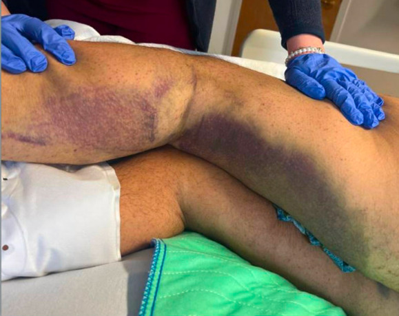
A 53-year-old man was admitted with progressive proximal muscle weakness and near-syncope. Review of systems was notable for pain and swelling of the left knee and ankle, a rash involving the lower extremities, and painful teeth and gums. His social history was significant for food insecurity, with a diet consisting predominantly of fast food and pizza. He reported consuming one to two alcoholic beverages daily and denied illicit drug use. Vital signs were within normal limits. Physical examination demonstrated large spontaneous ecchymoses and petechiae over the lower extremities (Figure 1), along with effusions and tenderness of the left knee and left ankle. Mild gingival bleeding and proximal lower extremity weakness were also present.
Initial laboratory evaluation revealed an elevated C-reactive protein (CRP) level of 56 mg/L (reference range, 0–3 mg/L), anemia with a hemoglobin of 9.6 g/dL (reference range, 14–17.2 g/dL), and folate deficiency (3.50 ng/mL; reference, >5.38 ng/mL). White blood cell count, platelet count, creatinine, liver transaminases, creatine kinase, vitamin B12, erythrocyte sedimentation rate (ESR), prothrombin time (PT), and partial thromboplastin time (PTT) were all within normal limits. Testing for human immunodeficiency virus (HIV) and syphilis was negative. Computed tomography (CT) of the chest, abdomen, and pelvis with intravenous contrast revealed no acute abnormalities. Magnetic resonance imaging (MRI) of the cervical, thoracic, and lumbar spine with and without contrast demonstrated only mild degenerative disc disease, without evidence of myelopathy or an underlying mass lesion. Electromyography was considered but was not immediately available.
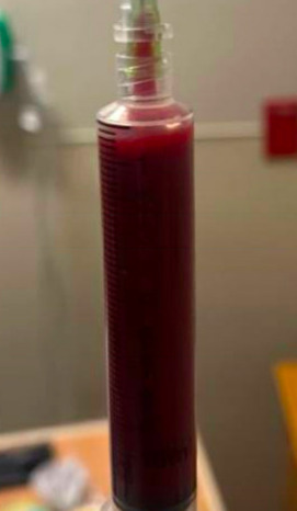
The combination of elevated C-reactive protein (CRP), petechial rash, joint swelling, and proximal muscle weakness initially raised concern for a systemic autoimmune or inflammatory disorder, prompting rheumatology consultation. Serologic testing, including antinuclear antibody (ANA), anti-SSA antibody, antineutrophil cytoplasmic antibody (ANCA), anti-tissue transglutaminase IgA antibody, rheumatoid factor (RF), and myositis-specific antibodies, was negative. Diagnostic arthrocentesis of the left knee yielded 15 mL of frankly bloody synovial fluid (Figure 2), prompting a shift in the differential diagnosis away from inflammatory disorders and toward causes of a bleeding diathesis. Subsequent testing revealed an undetectable serum vitamin C level (reference range, 0.2–2.1 mg/dL). The patient was started on oral ascorbic acid 1,000 mg daily, with progressive clinical improvement, and was discharged to a short-term rehabilitation facility.

The clinical manifestations of vitamin C (ascorbic acid) deficiency, or scurvy, have been recognized since at least the 16th century. The recommended daily intake of vitamin C is 75–90 mg, with fruits and green vegetables serving as the primary dietary sources. In resource-rich settings, scurvy most commonly occurs in individuals with neurodevelopmental disorders, neurocognitive impairment, eating disorders, or substance use disorders. However, those with restrictive diets or food insecurity are also at increased risk. The characteristic mucocutaneous manifestations of scurvy arise from impaired collagen synthesis and loss of capillary integrity rather than abnormalities of platelets or coagulation factors. Common findings include gingival bleeding, spontaneous ecchymoses, purpura, and perifollicular petechiae. Patients may also develop fatigue, anorexia, depression, neuromuscular weakness, and vasomotor instability, likely reflecting vitamin C’s essential role in fatty acid metabolism and cellular energy production.
Musculoskeletal manifestations are well described but may be underrecognized in adults. Typical features include arthralgias, myalgias, muscle weakness, and joint swelling, with symptoms often resulting from subperiosteal hemorrhage caused by capillary fragility. In severe cases, these manifestations can lead to marked functional impairment and mimic inflammatory arthritis, vasculitis, inflammatory myopathy, or neurologic disease. Hemarthrosis, however, is an uncommon manifestation in adults and has been reported only rarely.1–4 In this patient, recurrent falls likely contributed to the development of hemarthrosis by precipitating bleeding into a joint already predisposed by vascular fragility. If left untreated, scurvy can progress to life-threatening complications, including cardiovascular collapse and death. Treatment consists of oral ascorbic acid at a dose of 300-1000 milligrams daily for one month. Symptomatic improvement may be noted within 24 hours, and mucocutaneous abnormalities typically resolve within weeks. Our patient followed up with his primary care provider and neurologist within one month and noted improved energy and mobility, as well as the resolution of skin lesions.
From a clinical reasoning perspective, the identification of hemarthrosis proved to be the pivotal finding in this case. The patient initially presented with an unexplained multisystem illness characterized by weakness, elevated CRP, petechiae, ecchymoses, and joint swelling, raising concern for a systemic rheumatologic process such as idiopathic inflammatory myopathy, inflammatory arthritis, or vasculitis. However, the presence of frank blood on arthrocentesis fundamentally shifted the diagnostic approach and redirected the differential diagnosis. Hemarthrosis is highly unusual in rheumatologic disorders; joint effusion in inflammatory arthritis invariably yields yellow, cloudy synovial fluid with an elevated white blood cell count and relatively few red blood cells. In considering causes of bleeding diathesis, we noted that the patient had no thrombocytopenia, nor a predisposition to platelet dysfunction such as end-stage renal or liver disease. The patient’s history was not suggestive of an inherited bleeding disorder, such as von Willebrand disease or hemophilia. Additionally, normal prothrombin time (PT) and partial thromboplastin time (PTT) made an acquired coagulation factor deficiency unlikely. These findings narrowed the differential diagnosis to nutritional deficiencies as the most likely cause of the bleeding diathesis, prompting measurement of a serum vitamin C level. Although the elevated C-reactive protein (CRP) initially raised suspicion for an underlying inflammatory process, this case highlights that inflammatory markers are nonspecific and should be interpreted within the broader clinical context. Elevated CRP has previously been reported in scurvy and may reflect tissue injury and hemorrhage rather than autoimmunity.5
This case is also notable because scurvy developed in an individual without behavioral or psychiatric conditions typically associated with nutritional deficiencies. Instead, the primary risk factor was food insecurity, which led to a prolonged diet largely consisting of calorie-dense, micronutrient-poor foods, substantially increasing the risk of vitamin C deficiency. This case highlights the importance of obtaining a thorough dietary history as part of the social history, as nutritional factors may be easily overlooked yet prove critical to establishing the correct diagnosis. Screening for food insecurity may help identify patients at risk for nutritional deficiencies before severe clinical manifestations occur. Focused, nonjudgmental questions such as, “Can you tell me how many times you ate fruits or vegetables in the past week?” may provide more meaningful information than broader or potentially stigmatizing questions such as, “Do you get enough to eat?” In summary, scurvy should remain in the differential diagnosis for patients presenting with otherwise unexplained petechiae, ecchymoses, gingival bleeding, musculoskeletal pain, weakness, joint effusions, or hemarthrosis, particularly in the setting of dietary restriction or food insecurity.
Disclosures/Conflicts of Interest
None
Corresponding author
Heinrich-Karl Greenblatt, MD
Division of Rheumatology, SouthCoast Health
Division of Rheumatology, Roger Wiliams Medical Center
Email: hgreenblatt2019@gmail.com