

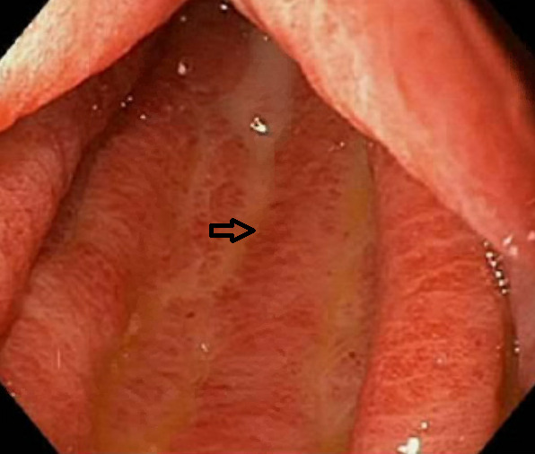
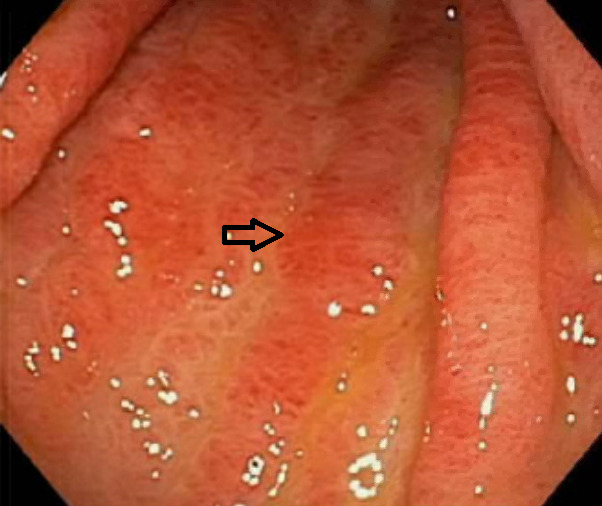
Gastric antral vascular ectasia (GAVE) is a rare but clinically important cause of chronic gastrointestinal bleeding.1–4 It can be associated with many systemic illnesses (cirrhosis, renal failure) including autoimmune diseases such as limited systemic sclerosis.2,3 Visible columns of red and ectatic vessels along the longitudinal folds of the antrum seen on endoscopy are pathognomonic for GAVE, also described as ‘watermelon stomach.’1–4 A 78-year-old woman with limited systemic sclerosis presented with iron deficiency anemia and melanotic stools. She initially underwent esophagogastroduodenoscopy (EGD) and colonoscopy which did not demonstrate abnormal findings. Video capsule endocopy was showed moderate GAVE as shown in Figures 1 and 2 (arrows showing vascular columns), which was successfully coagulated for hemostasis using an argon beam laser on a subsequent EGD.
Corresponding author
Maun R. Baral, MD
Resident Physician
Maun.rbaral@gmail.com
ORCiD: 0000-0002-5810-6751
Tel: 203-837-6079
Danbury Hospital,
24 Hospital Avenue
Danbury, CT, 06810
Author Conflicts of Interest
No disclosures or conflict of interest