Introduction
The most common vascular etiology of abdominal pain is mesenteric ischemia from an arterial embolism, arterial or venous thrombosis, and non-occlusive ischemia secondary to intestinal hypoperfusion. Furthermore, both arterial and venous occlusion can lead to intestinal ischemia from volvulus around the mesenteric defect. Acute mesenteric ischemia may also be observed in the setting of an underlying vasculitis; however, it may be difficult to determine whether arterial occlusion or spasm-related hypoperfusion is the cause of segmental intestinal infarction. Anterior nutcracker syndrome (NCS) occurs when the left renal vein is compressed between the abdominal aorta and the superior mesenteric artery (SMA) and is a rare cause of vascular abdominal pain. Posterior nutcracker syndrome is less common and occurs when the left renal vein is compressed between the abdominal aorta and a lumbar vertebral body.1–3 Renal nutcracker syndrome remains underdiagnosed with a poorly defined clinical course.4
Case
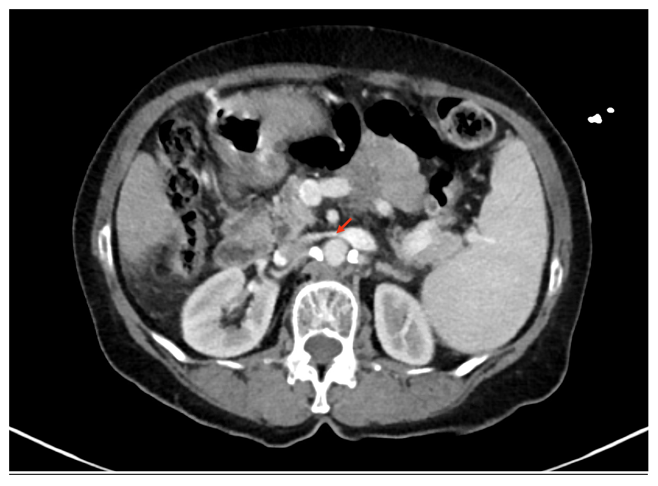
A 71-year-old woman who presented to hospital with sudden-onset intense abdominal pain over the lower abdomen (right iliac region, hypogastric region and left iliac region) with radiation to the left flank region. She also reported nausea, non-bloody & non-bilious vomiting, and loss of appetite. She denied hematemesis, melena, bright red blood per rectum and back pain. Her past medical history included asthma, hypertension, hyperlipidemia, type 2 diabetes mellitus, liver cirrhosis associated with non-alcoholic steatohepatitis and iron-deficiency anemia. Her medications at the time of admission were albuterol, rosuvastatin, empagliflozin, glimepiride, sitagliptan, furosemide, spironolactone, nadolol and omeprazole. On physical examination, her BMI was 20.45 kg/m². Vital signs were within normal limits and she was afebrile. She appeared to be in severe pain and was writhing in the hospital bed due to her discomfort. Her abdomen was soft and diffusely tender to palpation without guarding or rigidity. She had no back tenderness or costovertebral angle tenderness. Capillary refill time was <2 seconds on all 4 extremities. Radial, ulnar, brachial, subclavian and carotid pulses were 3+. There were no carotid bruits on auscultation. There was no radial-radial delay. Her femoral, popliteal, dorsalis pedis and posterior tibial pulses were also all 3+. There was no evidence of venous stasis and neuro-sensory examination was intact. Laboratory blood analysis was significant for serum hemoglobin 9.8 g/dL (11.7 - 15.0 g/dL), serum creatinine 1.52 mg/dL (0.50 - 1.10 mg/dL), serum lipase 128 U/L (8 - 78 U/L) and serum gamma-glutamyl transferase 124 U/L (8 - 35 U/L) with otherwise normal liver function tests. Microscopic examination of the urine showed evidence of hematuria. Abdominal imaging with a computed tomography (CT) scan of the abdomen & pelvis with contrast showed left renal vein narrowing as it crosses between the superior mesenteric artery and the aorta (Figure 1). There was no evidence of abdominal ascites. Anterior nutcracker syndrome of the left renal vein was diagnosed. The patient underwent a venogram study alongside intravascular ultrasound and was treated with left renal vein wall stenting (Boston Sci Wallstent 18 mm x 60 mm) and venoplasty. She tolerated the procedure well and was successfully discharged on systemic anticoagulation with rivaroxaban 20 mg daily for one month with close follow-up.

Discussion
Nutcracker phenomenon (NCP), also known as left renal vein entrapment, is characterized by obstructed outflow from the left renal vein (LRV) into the inferior vena cava (IVC) due to extrinsic LRV compression. The term “nutcracker” is used to describe these anatomic findings as this compression of the LRV is akin to a nutcracker crushing a nut. Nutcracker syndrome describes patients with clinical symptoms associated with nutcracker anatomy. Although the terms nutcracker phenomenon and nutcracker syndrome are frequently used interchangeably, nutcracker anatomy is not always associated with clinical symptoms and may represent a normal variant. The classical presentation of NCS usually includes hematuria associated with left flank pain. Patients may present with a wide variety of symptoms including varicocele, pelvic congestion syndrome and orthostatic proteinuria.5 The prevalence of NCS is unknown, however it has been described as more common in women.6–8 Anterior NCS results from compression of the LRV between the SMA and the aorta, while posterior NCS results from compression of the LRV between the aorta and the lumbar spine. Furthermore, conditions associated with an increased risk of NCS include retroperitoneal tumors, para-aortic lymphadenopathy, abdominal aortic aneurysm, left renal vein duplication, nephroptosis, lordosis and decreased retroperitoneal and mesenteric adipose tissue.9 Diagnostic tools commonly used to identify NCS include CT, magnetic resonance imaging (MRI) and Doppler ultrasonographic assessment of left renal vein diameter and peak velocity.6,8,10 Venography used to be the gold standard to confirm the diagnosis, but it is invasive and is now reserved for treatment purposes. During venography, the pressure gradient between the IVC and the LRV is measured, which should be > 3 mm Hg in NCS (normal pressure gradient is 0-1 mm Hg).6 Management of nutcracker syndrome remains controversial. Patients with mild symptoms are usually managed conservatively with regular surveillance and a focus on weight gain to increase retroperitoneal adiposity, leading to decompression of the left renal vein due to adjustment in position of the left kidney. If symptoms such as significant hematuria and left flank pain persist, such as in our patient, intervention with endovascular stenting or surgery may be necessary.11,12 In patients treated surgically with open repair, excellent long-term outcomes have been reported. Less-invasive endovascular stent placement appears to offer good results in the short term and long-term outcomes are still being investigated. Advances in venous stent technology are anticipated and may result in endovascular treatment of additional cases in the future.13
Author Contribution
All Authors (KVR, MR, GA, SV, DK, SVR) have reviewed the final manuscript prior to submission. All the authors have contributed significantly to the manuscript, per the ICJME criteria of authorship.
-
Substantial contributions to the conception or design of the work; or the acquisition, analysis, or interpretation of data for the work; AND
-
Drafting the work or revising it critically for important intellectual content; AND
-
Final approval of the version to be published; AND
-
Agreement to be accountable for all aspects of the work in ensuring that questions related to
Disclosures/Conflicts of Interest
None
Funding Information
None
Corresponding author
Karishma Vijay Rupani, MD MMSc
Resident Physician in Internal Medicine
Department of Medicine
Mount Sinai Beth Israel
350 East 17th Street
20th Floor
New York, NY 10003
karishma.rupani@mountsinai.org