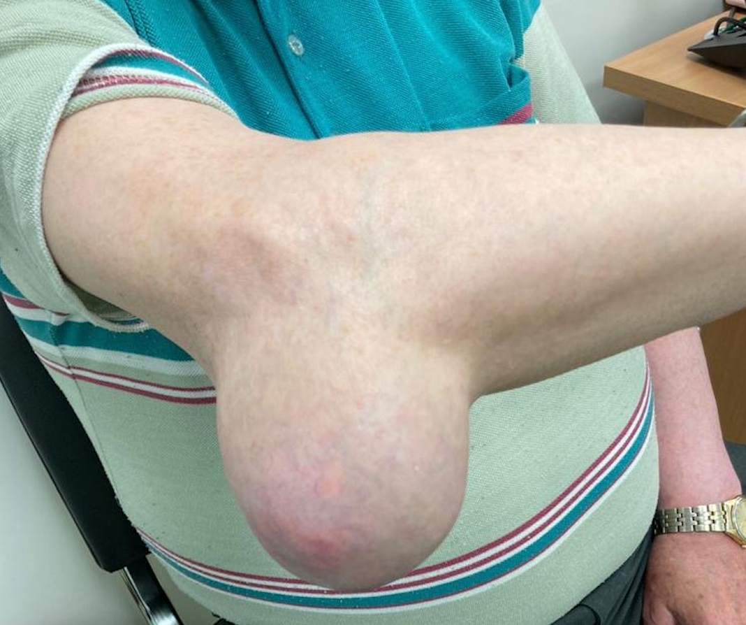
A 77-year-old man with a history of heart disease and hypertension presented to an outpatient plastic surgery department with a large, soft, painless, subcutaneous swelling on the extensor surface of his right elbow (Figure 1). The swelling had increased over ten years, with rapid growth in the preceding year. He denied any restriction in the movement of his elbow and reported no episodes of erythema or pain. Instead, he complained of functional limitations related to finding well-fitting clothing. He had no history of swelling or pain in other joints or tendons, and careful examination did not reveal deformity or tophi in other anatomical sites. He had no previous diagnosis of gout. His medications included a thiazide diuretic. There was no history of diabetes mellitus, kidney disease, excess alcohol intake, or active malignancy.

Magnetic resonance imaging showed a mass with T1 hyperintensity, heterogeneity, and several low signal foci. The mass was excised. Histology revealed multi-nodular amorphous eosinophilic material with a surrounding histiocytic giant cell reaction. Radiological and histological features were in keeping with gouty tophi. The patient recovered without complications. His serum urate levels were 490mmol/L. He was commenced on urate-lowering therapy and was symptom-free at the last clinical follow-up.
Gout or monosodium urate crystal deposition disease is initiated by plasma urate concentrations that exceed the limit of urate solubility, typically greater than 400 mmol/L (6.8mg/dL). Urate is produced in the body during the metabolism of purines. This production is balanced by renal and extra-renal, predominantly intestinal, elimination. Over production or decreased elimination leads to hyperuricemia.1 Several interacting checkpoints, including the tendency of formation and deposition of urate crystals and the host inflammatory response, are required to produce the clinical manifestations of gout.2 Pseudotumor has been reported elsewhere in the elbow, knee, ankle, and soft tissue sites, although usually in patients with an established diagnosis of gout.3–6 Gout should remain a differential diagnosis for peri-articular or tendinous masses, even without a prior diagnosis. Risk factors for gout should alert one to the possibility of occult disease and should prompt investigation and treatment to prevent ongoing complications.
AUTHOR CONTRIBUTIONS
All authors have reviewed the final manuscript prior to submission. All the authors have contributed significantly to the manuscript, per the International Committee of Medical Journal Editors criteria of authorship.
- Substantial contributions to the conception or design of the work; or the acquisition, analysis, or interpretation of data for the work; AND
- Drafting the work or revising it critically for important intellectual content; AND
- Final approval of the version to be published; AND
- Agreement to be accountable for all aspects of the work in ensuring that questions related to the accuracy or integrity of any part of the work are appropriately investigated and resolved.
CONFLICTS OF INTEREST
The authors have no conflicts of interest to disclose.