Background
The lymphatic system functions to absorb and transport chyle from peripheral lymphatics and the cisterna chyli into the systemic circulation. Chyle is a white, milky fluid produced during the digestion of fatty meals in intestinal lacteal cells. It is primarily composed of triglycerides, proteins, and lymphocytes, predominantly T cells; therefore, chylous leaks represent a complex physiological and anatomical challenge.1 Chylous loss can lead to malnutrition and immunosuppression, contributing to significant morbidity and mortality. Reported mortality rates reach 40–70% in chylous ascites and up to 82% 90-day mortality in chylothorax.2,3
Peritoneal dialysis involves instilling clear dialysate into the peritoneal cavity. Despite a declining incidence, peritonitis remains a major complication and a leading cause of morbidity, mortality, and peritoneal dialysis modality failure in peritoneal dialysis; therefore, it must always be excluded.4 Any change in the color or consistency of the dialysate warrants prompt evaluation and fluid analysis to establish the diagnosis. In this report, we present a case of chylous ascites resulting from lymphatic injury during dialysis catheter insertion, which resolved with conservative management alone.
Case Presentation
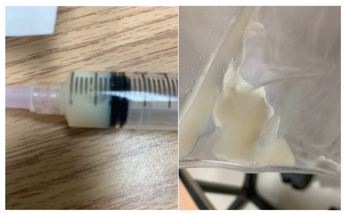
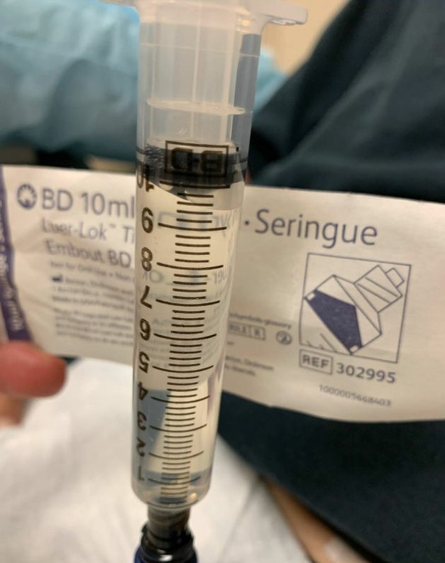
A 72-year-old male with a history of progressive type 2 diabetes mellitus, hypertension, pulmonary fibrosis, and stage IV chronic kidney disease complicated by uremia underwent placement of a peritoneal dialysis (PD) catheter by laparoscopy, which was uncomplicated. Operative notes documented laparoscopic placement using trocars in the left upper quadrant, epigastrium, and left mid-abdomen. The catheter was introduced above the umbilicus over the right rectus abdominis, with the deep cuff seated in the rectus sheath, tunneled across the midline, and anchored in the pelvis to the right medial umbilical ligament, with good flow achieved. Twenty days after uneventful placement, he presented for initial flush and training. After a normal initial flush, the subsequent fluid drained was noted to be very thick and milky white in appearance (Figure 1).
He had no history of liver cirrhosis, malignancy, abdominal surgery, or trauma and reported only occasional crampy “gas-like” discomfort after surgery, which had largely passed. He denied current fever, chills, abdominal pain, nausea, or vomiting. Physical examination revealed a soft, non-tender abdomen without guarding or rigidity. The catheter insertion site was clean, without erythema or purulence. The appearance in combination with the lack of symptoms raised suspicion for chylous ascites. Analysis of the peritoneal dialysis effluent demonstrated albumin <1 g/dL and total protein <2 g/dL. Lactate dehydrogenase was 64 U/L. Amylase and lipase levels were 12 U/L and 8 U/L, respectively. Cholesterol was 45 mg/dL, and triglycerides were elevated at 653 mg/dL. Red blood cells were <3000/µL, and nucleated cells were 863/µL. Fluid cytology revealed predominantly macrophages with numerous eosinophils and few neutrophils. The differential cell count showed 2% neutrophils, 8% lymphocytes, 41% eosinophils, and 47% monocytes/macrophages, corresponding to an absolute neutrophil count of approximately 17 cells/µL. Cultures were negative for bacterial and fungal growth. Cytology was negative for malignant cells. Glucose was markedly elevated at 1266 mg/dL, attributed to peritoneal dialysis fluid flush. Serum CA-125 was within normal limits at 12 U/mL. Chylous ascites was strongly suspected, and antibiotics were not prescribed.
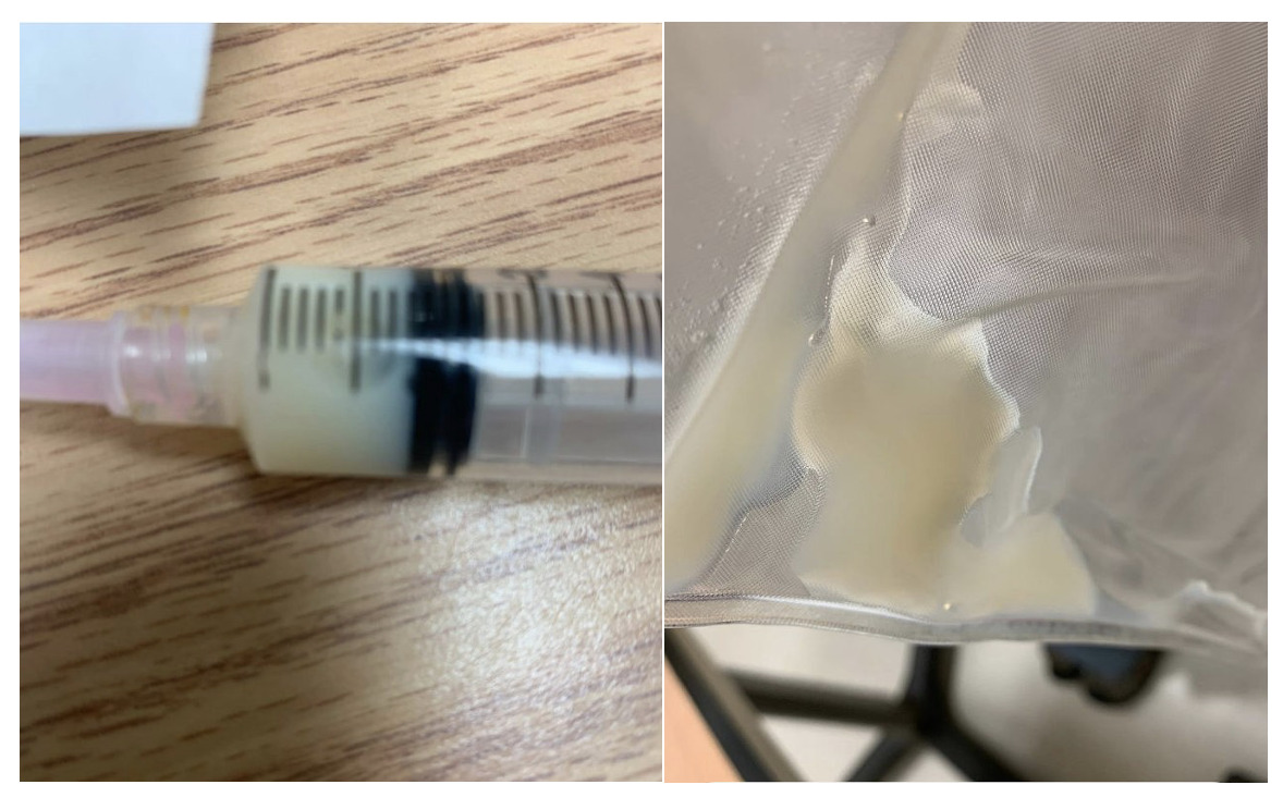
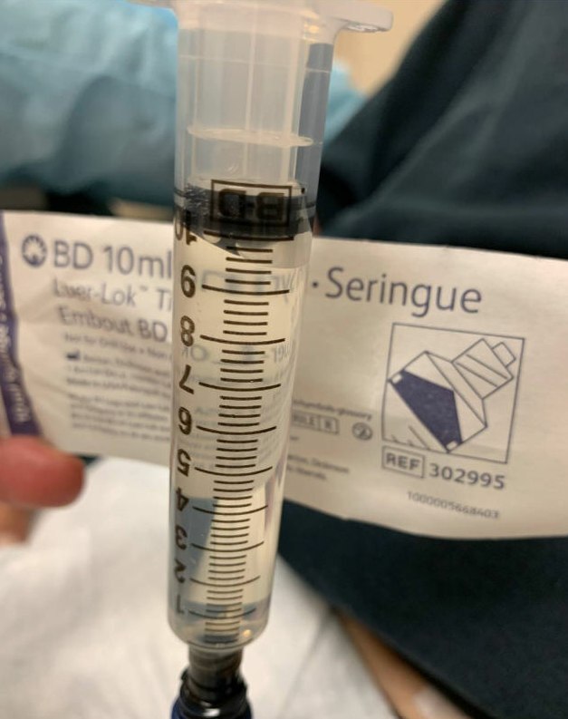
A repeat ascitic fluid analysis two weeks later continued to show milky fluid, with triglycerides further increased to 874 mg/dL and nucleated cells 2072/µL. Glucose decreased to 320 mg/dL. Gram stain and cultures remained negative. Non-contrast computed tomography of the abdomen and pelvis, obtained without dialysate dwell, demonstrated the peritoneal dialysis catheter in appropriate position, with a small volume of free fluid in the pelvis and no abdominal or pelvic lymphadenopathy, lymphatic obstruction, or other imaging evidence of lymphatic injury or a secondary cause of chylous ascites. Given the patient’s stable clinical status, absence of infectious signs, and negative microbiologic studies, a diagnosis of chylous ascites was confirmed. No antibiotics were administered, and peritoneal dialysis was continued with close monitoring. At one-month follow-up, the peritoneal dialysis effluent had become clear with resolution of the chylous appearance (Figure 2). Due to concern for possible drug-related chylous ascites associated with calcium channel blockers, amlodipine was discontinued at follow-up; however, the dialysate had already cleared prior to cessation of the medication.
Discussion
Chylous ascites is a known but rare complication of peritoneal dialysis. It is important to distinguish it clinically, as it can mimic peritonitis and lead to unnecessary antibiotic use. Although cloudy dialysate is commonly associated with peritoneal dialysis–related peritonitis, truly milky effluent is uncommon and should raise suspicion for chylous ascites. According to the 2022 ISPD peritonitis guidelines, the diagnosis of peritonitis requires at least two of the following: compatible clinical features (abdominal pain or cloudy effluent); a peritoneal fluid white cell count greater than 100/mm3 (0.1 × 10⁹/L) after a dwell time of at least two hours, with more than 50% neutrophils; or a positive effluent culture [19]. In our patient, there was no abdominal pain or systemic symptoms, cultures were persistently negative, and the neutrophil percentage was well below this threshold, making peritonitis unlikely despite the cloudy appearance of the effluent.
The etiology of primary chylous ascites is most commonly due to lymphatic abnormalities, including rupture, trauma, or obstruction, which increase intralymphatic pressure.5 Secondary causes in adults are most frequently associated with cirrhosis and malignancy, particularly lymphoma, whereas in children, it is more commonly related to congenital defects.6<u>,7</u> Additional causes include trauma (iatrogenic and non-iatrogenic), infections, inflammatory conditions, cardiac disease (e.g., right heart failure), gastrointestinal disorders, and drug-related etiologies.6 Calcium channel blockers have been associated with chylous ascites, although the mechanism remains unclear.7 In a systematic review and meta-analysis evaluating this association, most patients were undergoing peritoneal dialysis. A total of 48 cases were included. Symptoms such as abdominal distension and/or pain, along with chylous ascites, developed within 2–3 days of initiating therapy and resolved within 24 hours of discontinuation. Rechallenge was performed in 10 patients, all of whom experienced recurrence of chylous effluent, which again resolved within 24 hours after stopping the medication.8 These findings highlight the importance of recognizing this association, particularly in patients with impaired kidney function and those undergoing peritoneal dialysis. In our case, the calcium channel blocker was discontinued; however, the dialysate had already begun to clear prior to its discontinuation.
For diagnosis, abdominal paracentesis—or, in this case, peritoneal dialysate analysis—is the initial step. Chylous fluid typically appears milky and cloudy, in contrast to the straw-colored fluid seen in ascites related to cirrhosis or portal hypertension. In some cirrhotic cases, the fluid may appear opalescent due to malignancy or infection without elevated triglyceride levels.9 Triglyceride levels above 200 mg/dL generally confirm the diagnosis, although a cutoff of 110 mg/dL has also been proposed.10–13 Management of chylous ascites requires a multidisciplinary, stepwise approach based on the underlying etiology and the patient’s nutritional status. Conservative therapy should be the first-line approach. In low-output states, enteral nutrition with a low-fat, high-protein diet enriched with medium-chain triglycerides is preferred, as these are absorbed directly into the portal circulation rather than the lymphatic system.3 Somatostatin analogues may also be considered, as they reduce lymphatic flow.14 If conservative measures fail, additional interventions such as lymphangiography with embolization may be used to localize and treat the leak. Surgical exploration is reserved for rare cases with high-output leaks refractory to conservative therapy.
In a similar report, a 52-year-old woman with cystic fibrosis status post lung transplantation and end-stage renal disease on peritoneal dialysis developed milky dialysate during hospitalization for pneumonia. Fluid triglyceride levels were 274 mg/dL, confirming chylous ascites. Imaging showed no lymphatic obstruction. Conservative dietary management was attempted; however, due to persistent symptoms and ongoing chylous effluent, she was transitioned to hemodialysis, which resulted in resolution.15 Another notable case involved a 78-year-old patient on peritoneal dialysis who developed chylous ascites (triglycerides 149 mg/dL) following accidental infusion of overheated dialysate, later complicated by bacterial peritonitis. Common etiologies—including malignancy, cirrhosis, tuberculosis, pancreatitis, and calcium channel blocker use—were excluded. Thermal injury was thought to cause intestinal barrier dysfunction, bacterial translocation, and lymphatic damage, leading to delayed inflammation and chylous ascites.16
Conclusion
Chylous ascites in peritoneal dialysis patients remains an uncommon but clinically significant entity that may be mistaken for peritonitis. Prompt recognition and appropriate fluid analysis are critical to avoid unnecessary antibiotic therapy. After exclusion of secondary causes, catheter-related lymphatic injury should be considered. This case demonstrates that in stable patients, conservative management alone can be effective, supporting a stepwise, individualized treatment approach.
Conflicts of Interest/Disclosures
None
Corresponding author
Huda Salih, MD
Department of Internal Medicine,
Rochester General Hospital, Rochester, NY
Email: hudafaisal1996@gmail.com