Background
Scorpion venoms contain neurotoxic peptides that affect voltage-gated ion channels and can trigger exaggerated sympathetic and parasympathetic neurotransmission. Most stings cause local pain, paresthesia, and self-limited symptoms; however, systemic envenomation can produce cardiovascular, pulmonary, neurologic, metabolic, and gastrointestinal complications.1,2
Acute pancreatitis following scorpion envenomation has been described primarily in regions with medically significant species, including Tityus and Leiurus, but reports from the United States are rare.3,4 Georgia has several native or regionally documented scorpions, including Vaejovis carolinianus and Centruroides hentzi.5,6 Because the scorpion implicated in this case was not captured, its species cannot be confirmed. This case is educational for hospital providers because it links a plausible environmental exposure to a severe abdominal presentation, while emphasizing the need to maintain a broad differential for pancreatitis and avoid overattribution when evidence of exposure is incomplete. C. hentzi has been characterized as a comparatively low-risk member of a medically important genus, despite diverse venom proteins.7
Case Presentation
A 26-year-old non-verbal man with autistic disorder was admitted to our facility after transfer from an outside hospital for acute pancreatitis. His available past medical history was notable for autism spectrum disorder with nonverbal baseline status. Additional history, including baseline functional status, home medications, recent antibiotic exposure, supplements, alcohol exposure, and prior gastrointestinal symptoms, was obtained from his parents and available records. No medication, recent antibiotic exposure, supplement, alcohol use, trauma, or other clear pancreatitis trigger was identified in the available documentation. History was provided by his parents, who reported three days of abdominal pain, posturing, drooling, vomiting, and visible distress. They also reported a high prevalence of scorpions in the home, although no direct witness to a sting or captured scorpion was available. The family also reported an approximately 30-pound weight loss over the preceding six months. This weight loss was clinically significant because it broadened the differential diagnosis to include reduced intake from gastroparesis, chronic gastrointestinal disease, obstructive pathology, inflammatory disease, and malignancy. No gastric outlet obstruction or pancreaticobiliary mass was identified on available imaging or endoscopic evaluation; however, the weight loss was not assumed to be fully explained by the acute episode alone and outpatient gastroenterology follow-up was arranged.
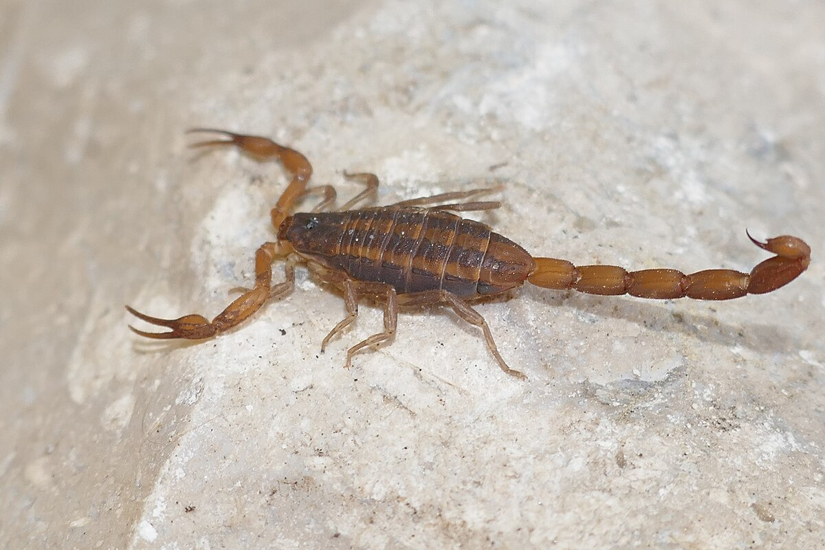
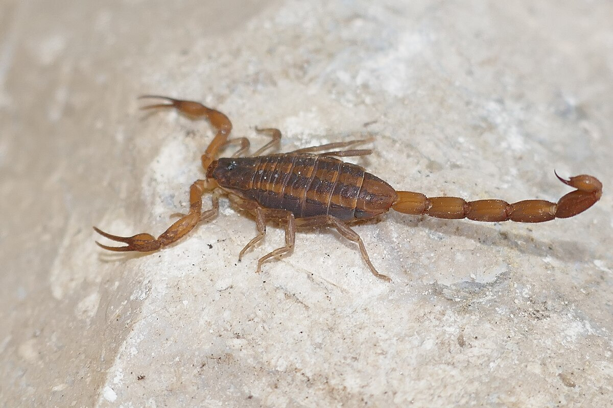
On presentation, the patient was afebrile and hemodynamically stable. Physical examination showed severe epigastric tenderness with radiation to the back and significant abdominal distension. A localized area of left flank swelling was noted during the hospitalization. A soft-tissue ultrasound of the area showed no acute abnormality. The family also reported taking a photograph of a scorpion found in the home, as shown in Figure 1, although the scorpion was not captured and its species was not confirmed. Evaluation for common causes of acute pancreatitis was unrevealing. Liver enzymes were within normal limits, and CT (contrast-enhanced computed tomography) imaging did not show gallstones, biliary dilation, or gallbladder abnormality. Triglycerides were 76 mg/dL, serum calcium was within normal limits, and renal function was normal. There was no documented alcohol use, medication trigger, traumatic injury, or obstructing pancreaticobiliary lesion in the available records. Immunoglobulin testing was unrevealing except for a low level of IgG subclass 3. Key laboratory values are summarized in Table 1.
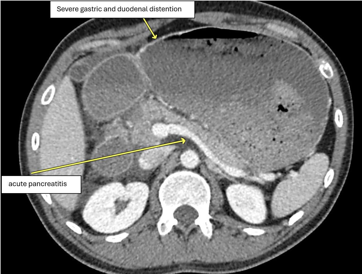
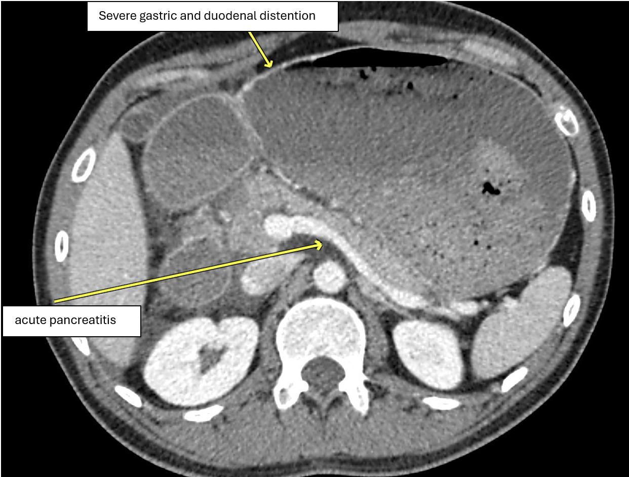
A contrast-enhanced CT of the abdomen and pelvis showed pancreatic head edema with mild peripancreatic fat stranding and free fluid, consistent with acute pancreatitis as shown in Figure 2. Follow-up noncontrast CT showed persistent pancreatic swelling and peripancreatic stranding with trace ascites, likely related to pancreatitis, as well as small bilateral pleural effusions with bibasilar compressive atelectasis or pneumonia. The previously severe gastric distension had resolved. Esophagogastroduodenoscopy (EGD) was subsequently performed because of persistent concern for impaired gastric emptying. EGD demonstrated gastroesophageal reflux disease (GERD), gastritis, duodenitis, and retained gastric food residue consistent with gastroparesis. No pyloric stenosis or gastric outlet obstruction was identified. The patient was managed with bowel rest, intravenous fluid resuscitation, correction of hypokalemia, and opioid analgesia as needed. A nasogastric tube was placed for intermittent suction and gastric decompression, although maintaining the tube was difficult because the patient repeatedly removed it. After clinical improvement, his diet was advanced cautiously, and he tolerated oral intake. His pancreatic enzymes decreased from an initial lipase of 877 U/L and amylase of 590 U/L to 266 U/L and 100 U/L, respectively. C-reactive protein also improved from >300 mg/L to 140 mg/dL. These laboratory trends paralleled the patient’s clinical improvement, including reduced abdominal distension, improved pain behaviors, tolerance of oral intake, and stabilization for discharge.
Before discharge, the care team reviewed return precautions with the patient’s parents, including recurrent vomiting, worsening abdominal pain or distension, fever, inability to tolerate oral intake, dehydration, or recurrent concern for envenomation. Given the reported high prevalence of scorpions in the home, the family was counseled to pursue environmental mitigation measures, including a home inspection, pest-control measures, sealing potential entry points, reducing indoor clutter, and avoiding direct handling of scorpions. Outpatient gastroenterology follow-up was arranged to monitor recovery, gastroparesis symptoms, nutritional status, and weight trajectory.
Discussion
This case describes acute pancreatitis with severe gastric and duodenal dilation after suspected scorpion envenomation. The diagnosis of acute pancreatitis was supported by characteristic epigastric pain radiating to the back, serum lipase and amylase elevations greater than three times the upper limit of normal, and CT findings consistent with pancreatitis.8 The clinical course was also notable for gastroparesis without mechanical gastric outlet obstruction.
The causal relationship remains uncertain and should therefore be interpreted cautiously. A scorpion was not captured, and the sting was not directly witnessed. Nevertheless, the household exposure history, left-flank swelling, absence of common etiologies in the provided evaluation, and compatible neurogastrointestinal physiology support the suspected envenomation as a plausible trigger. In future similar cases, specimen identification, toxicology consultation, serial pancreatic enzyme measurements, right upper quadrant ultrasonography or MRCP/EUS (Magnetic Resonance Cholangiopancreatography/Endoscopic Ultrasound) when indicated, medication review, and careful documentation of alcohol exposure, calcium, triglycerides, and biliary findings would strengthen etiologic attribution.9,10
Scorpion-induced pancreatitis is thought to arise from toxin-mediated autonomic dysregulation. Neurotoxins can trigger release of catecholamines and acetylcholine, producing a mixed sympathetic and parasympathetic storm.1,2 Proposed pancreatic mechanisms include sphincter of Oddi spasm, increased pancreatic exocrine secretion, pancreatic microvascular injury, and premature enzyme activation.3,4,11 The same autonomic imbalance may impair gastric motility, providing a biologically plausible explanation for concomitant gastroparesis and acute gastric dilation in this patient.
The geographic context is important. Severe systemic scorpion envenomation is more often reported from tropical and subtropical regions with highly toxic species.2,3 In the southeastern United States, severe systemic manifestations are uncommon, and C. hentzi is generally regarded as far less dangerous than more medically significant Centruroides species.5–7 However, venom composition, dose, host susceptibility, communication barriers, and delayed recognition may influence presentation. In this case, the patient’s non-verbal status and autistic disorder likely increased diagnostic complexity because symptom onset, pain localization, and exposure timing depended on caregiver observation. This case also illustrates practical strategies for evaluating serious abdominal pathology in a nonverbal patient. The diagnostic history depended on caregiver recognition of pain behaviors, including posturing, visible distress, drooling, vomiting, and changes from baseline behavior. The team reconstructed the symptom timeline with the parents, reviewed environmental exposures in the home, and incorporated the family-provided scorpion photograph while acknowledging that no witnessed sting or captured specimen was available. These steps were essential to generating the exposure hypothesis while maintaining a broad differential diagnosis.
Management remained supportive and aligned with current principles for acute pancreatitis: early assessment, fluid resuscitation, analgesia, correction of metabolic derangements, avoidance of unnecessary invasive interventions, and early oral feeding once clinically tolerated.9,10 Gastric decompression was appropriate for severe gastric dilation and vomiting, and EGD helped exclude pyloric stenosis or obstructing pathology when gastroparesis persisted. The patient’s improvement with supportive care is consistent with the usually transient course of toxin-mediated pancreatic and autonomic dysfunction described in prior reports.3,11 In regions where severe systemic scorpion envenomation is well recognized, antivenom therapy has been studied; however, these findings should not be extrapolated to an unconfirmed scorpion exposure in Georgia in the absence of severe cardiopulmonary instability.12
Conclusion
Acute pancreatitis with severe gastric dilation and gastroparesis may occur after suspected scorpion envenomation, even in regions where native species are usually considered to cause mild stings. This case underscores the value of caregiver history in non-verbal patients, careful exclusion of common pancreatitis etiologies, cautious species attribution, and supportive management directed at pancreatitis and its complications.
Conflicts of interest/Disclosures
The authors declare no conflicts of interest related to this case report.
Corresponding author
Parth Adrejiya, MD
Wellstar Spalding Regional Hospital, Griffin, Georgia, USA
Email: parthadrejiya@gmail.com