QUESTION 1: Do all patients with colonic ischemia require antibiotics?
A 46-year-old man with end-stage renal disease (ESRD) secondary to focal segmental glomerulosclerosis, on maintenance hemodialysis for the past six months, as well as hypertension and hyperlipidemia, presented with a one-day history of abrupt-onset left lower quadrant abdominal pain. The pain progressively worsened over several hours, and became associated with intermittent episodes of hematochezia. His home medications included low-dose aspirin, atorvastatin, amlodipine, sevelamer, lisinopril, and carvedilol.
On examination, he was afebrile with a blood pressure of 102/62 mmHg and a heart rate of 105 beats/minute. Cardiopulmonary examination was unremarkable, with no pericardial rub. Abdominal examination demonstrated a nondistended abdomen with mild left lower quadrant tenderness and voluntary guarding. Bowel sounds were normoactive. Laboratory studies revealed a white blood cell count of 15,500/µL, hemoglobin of 11.6 g/dL, blood urea nitrogen of 45 mg/dL, and serum creatinine of 4.6 mg/dL.
Computed tomography of the abdomen and pelvis without intravenous contrast demonstrated segmental wall thickening involving the distal transverse and descending colon. Gastroenterology was consulted, and colonoscopy revealed edematous, friable mucosa with ulceration and submucosal hemorrhage involving the distal transverse and descending colon, findings consistent with ischemic colitis. Notably, the patient had experienced a similar episode of ischemic colitis approximately 14 months earlier, which was managed successfully with conservative therapy consisting of bowel rest, cautious intravenous hydration, and analgesia. A diagnosis of ischemic colitis is made. He had a similar diagnosis approximately 14 months ago and was managed conservatively with bowel rest, gentle hydration and analgesics. How should he be managed now?
A: The American College of Gastroenterology (ACG) recommends risk stratification of colonic ischemia based on the presence or absence of clinical features associated with poor outcomes, as this approach guides management decisions. Established predictors of severe disease include male sex, hypotension (systolic blood pressure <90 mmHg), tachycardia (heart rate >100 beats/minute), abdominal pain without rectal bleeding, blood urea nitrogen (BUN) >20 mg/dL, hemoglobin <12 g/dL, lactate dehydrogenase (LDH) >350 U/L, and serum sodium <136 mEq/L. Chronic hemodialysis and poor Eastern Cooperative Oncology Group (ECOG) performance status are also independent predictors of severe disease and adverse outcomes.1–4
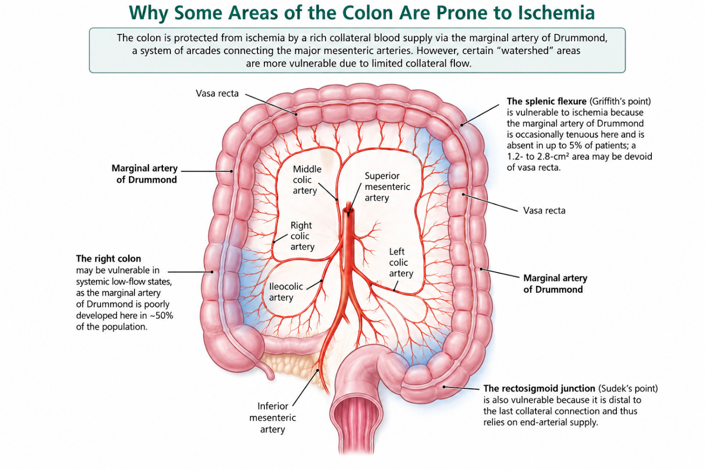
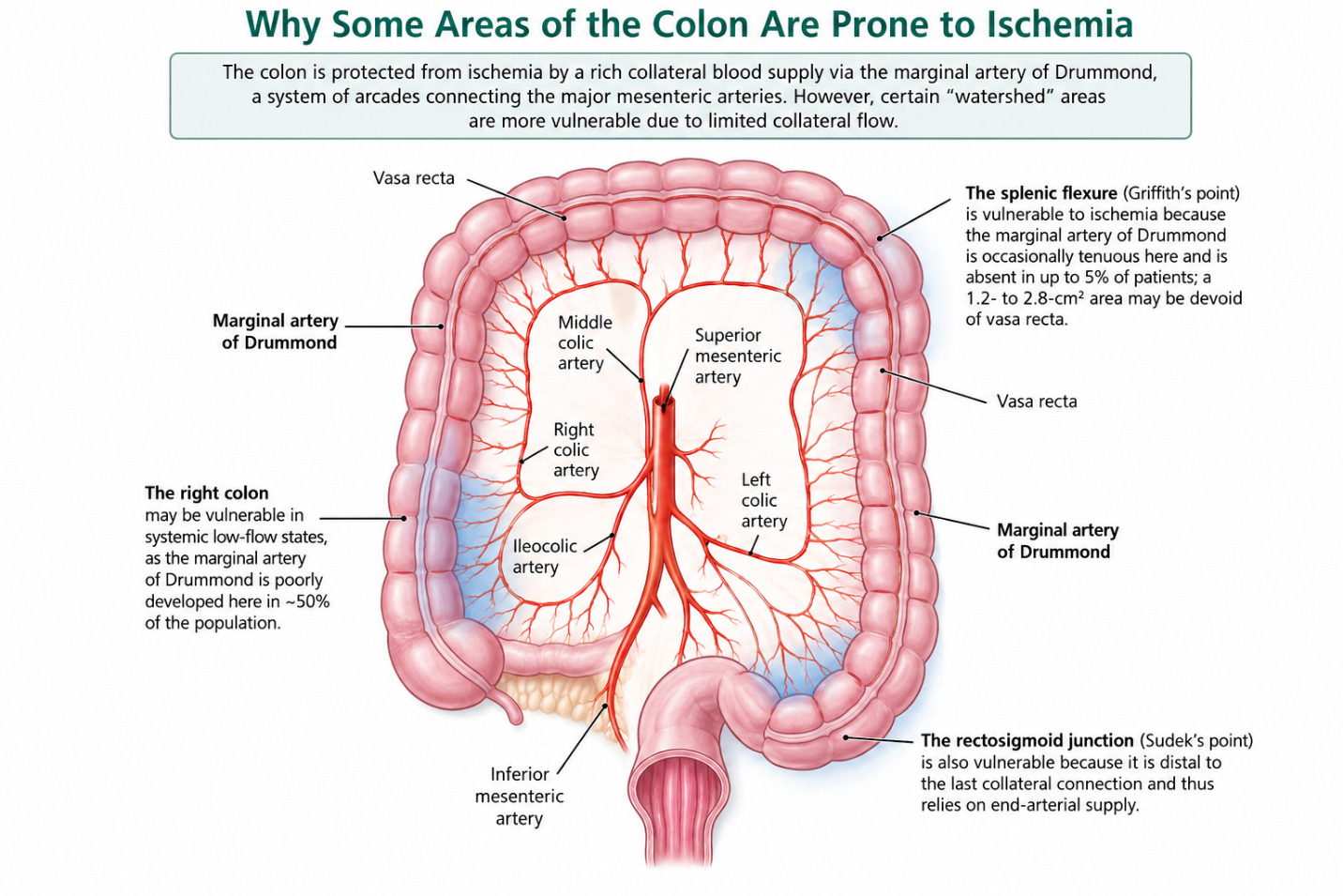
Colonic ischemia is classified as mild, moderate, or severe based on clinical presentation and risk profile (Figure 1). Mild colonic ischemia is characterized by typical symptoms with supportive findings on abdominal imaging or lower endoscopy, no risk factors for poor outcomes, and no evidence of peritonitis. Moderate colonic ischemia is defined by typical clinical and radiographic or endoscopic findings in the presence of up to three risk factors for poor outcomes, without peritoneal signs or an immediate indication for laparotomy. Severe colonic ischemia is diagnosed in patients with more than three risk factors for poor outcomes or the presence of any high-risk feature, including peritoneal signs on examination, pneumoperitoneum, pneumatosis intestinalis, portal venous gas on imaging, or gangrene identified during colonoscopy. Chronic hemodialysis and poor ECOG performance status further increase the likelihood of severe disease.1–4
Patients with at least moderate colonic ischemia, particularly those with more than two risk factors for poor outcomes, should receive broad-spectrum antibiotics in addition to supportive care and correction of potential precipitating factors. These patients require close inpatient monitoring and early surgical consultation to facilitate prompt laparotomy should clinical deterioration or evidence of bowel infarction develop.5 In this patient, the previous episode of colonic ischemia was most consistent with mild disease and occurred before the initiation of hemodialysis for end-stage renal disease. The subsequent development of dialysis dependence represents an important change in risk profile, as chronic hemodialysis is an independent predictor of severe colonic ischemia and poorer clinical outcomes.
QUESTION 2: How should physicians manage patients with jejunal diverticulitis?
A 68-year-old man with a history of gastroesophageal reflux disease presented with a 2-day history of progressively worsening left-sided abdominal pain associated with nausea and low-grade fever. He denied diarrhea, hematochezia, melena, dysuria, or hematuria. He had no history of alcohol or illicit drug use and no known family history of inflammatory bowel disease or gastrointestinal malignancy. On presentation, his temperature was 99.4°F, heart rate was in the 80s beats/min, and blood pressure was in the 110s/70s mmHg. Cardiopulmonary examination was unremarkable. The abdomen was mildly distended, with focal tenderness in the left upper quadrant. There was no guarding or rebound tenderness, and bowel sounds were normoactive. Laboratory evaluation demonstrated marked leukocytosis with a white blood cell count of 22.5 × 10³/μL. Hemoglobin was 14.1 g/dL and platelet count was 136 × 103/μL. Basic metabolic panel and liver function tests were within normal limits. Lipase was 13 U/L. Computed tomography (CT) of the abdomen and pelvis demonstrated findings consistent with jejunal diverticulitis. How should he be managed now?
A: Jejunal diverticulosis is an uncommon disorder of the small intestine and is considerably less frequent than colonic diverticulosis. Reported prevalence varies between approximately 0.5% and 6%, with a predilection for older adults, particularly men. Jejunal diverticula are usually acquired pseudodiverticula resulting from herniation of the mucosa and submucosa through the muscular layer of the intestinal wall. They typically arise along the mesenteric border of the small bowel, with the jejunum more commonly affected than the Ileum. Jejunal diverticula may occur in isolation or in association with diverticular disease involving other portions of the gastrointestinal tract, including the duodenum and colon.6,7
Patients with jejunal diverticulitis commonly present with abdominal pain, nausea, vomiting, and fever. The location of pain depends on the involved segment of jejunum but may be predominantly left-sided, as in our patient. Although most patients with jejunal diverticulosis remain asymptomatic, approximately 10%–20% develop complications. These include diverticulitis, abscess formation, gastrointestinal hemorrhage, obstruction, perforation, and peritonitis. Because of its rarity and nonspecific presentation, jejunal diverticulitis can be challenging to recognize clinically. CT of the abdomen and pelvis is the primary diagnostic modality. Typical CT findings include discrete, round or ovoid, air-, fluid-, or contrast-filled outpouchings arising from and extending beyond the expected bowel lumen. Other diagnostic modalities, including small-bowel follow-through and double-balloon enteroscopy, have been described. Enteroclysis can improve visualization through enhanced bowel distension and has historically been considered a useful technique for demonstrating small-bowel diverticula.6,7
There is no disease-specific Hinchey classification for small-bowel diverticulitis. However, the Hinchey classification developed for colonic diverticulitis is often applied by analogy to small-bowel disease, particularly when imaging demonstrates peritoneal involvement. Some centers have also used the modified Hinchey classification (stages 0–IV) to describe the severity of small-bowel diverticulitis (Tables 1 and 2).8,9
Management depends primarily on the severity of disease and the presence or absence of complications. Patients with uncomplicated jejunal diverticulitis can generally be managed conservatively with bowel rest, intravenous fluids, analgesia, and antibiotics when clinically indicated. Antibiotic therapy should provide coverage for enteric gram-negative and anaerobic organisms. In clinically stable patients without systemic toxicity, selected cases may be managed with oral antibiotics and close outpatient follow-up.10
Diverticular abscesses may occur in up to 16% of patients with acute diverticulitis. Management depends on abscess size and clinical severity. Small abscesses (<2 cm) may be managed with intravenous antibiotics alone, whereas larger or persistent abscesses may require image-guided percutaneous drainage or surgical intervention. Abscesses corresponding to Hinchey stages Ib and II can be successfully managed with percutaneous drainage in approximately 50% of cases. Patients who fail conservative management or develop large abscesses or perforation may require surgical intervention with diverticulectomy or segmental small-bowel resection. Our patient had modified Hinchey class 1a jejunal diverticulitis and responded well to conservative treatment with intravenous antibiotics and analgesia. His clinical improvement allowed transition to oral antibiotics and discharge without surgical intervention. Recognition of jejunal diverticulitis is important not only in patients presenting with abdominal pain but also in those with otherwise unexplained gastrointestinal bleeding.10
Disclosures/Conflicts of Interest
None
Corresponding Author
Kwame Dapaah-Afriyie, MD
Professor of Medicine, Clinical Educator
Warren Alpert Medical School at Brown University
Division Director
Division of Hospital Medicine
The Miriam Hospital, 164 Summit Avenue, Providence, RI 02906