Introduction
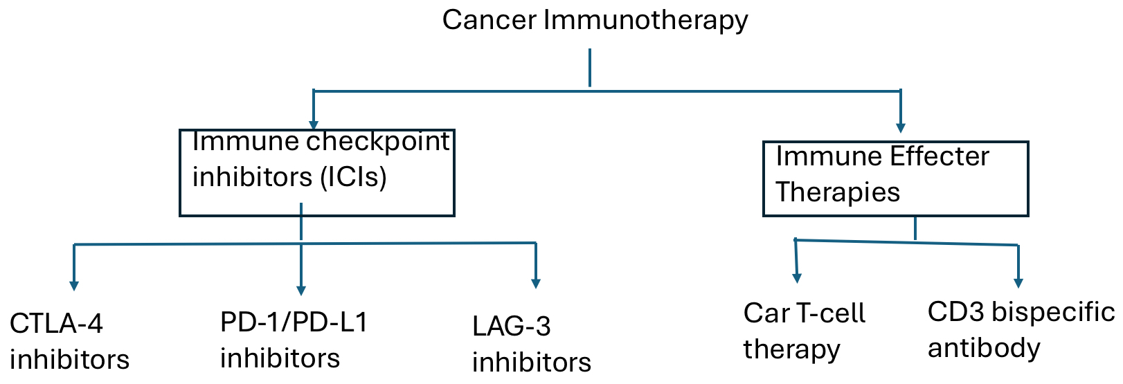
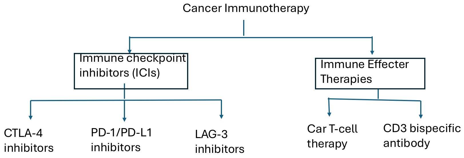
Immune checkpoint inhibitors (ICIs) and immune effector cell (IEC) therapies have transformed oncology. ICIs targeting cytotoxic T-lymphocyte–associated protein 4 (CTLA-4), programmed cell death protein 1 (PD-1), programmed death-ligand 1 (PD-L1), and lymphocyte activation gene-3 (LAG-3) demonstrate durable clinical benefit across multiple malignancies, including melanoma, non–small cell lung cancer, and renal cell carcinoma.1–5These agents regulate distinct phases of T-cell activation, and LAG-3 inhibition synergizes with PD-1 blockade to reverse T-cell exhaustion and enhance antitumor immunity.5,6 Concurrently, CD3-engaging bispecific antibodies and chimeric antigen receptor (CAR) T-cell therapies produce deep responses in relapsed or refractory hematologic malignancies, expanding immune-based cancer therapy.7–10 However, these advances are accompanied by immune-mediated toxicities resulting from dysregulated immune activation and loss of peripheral tolerance.11–13
Management of immune-related adverse events (irAEs) and IEC-associated toxicities is guided by frameworks from the American Society of Clinical Oncology (ASCO), the National Comprehensive Cancer Network (NCCN), and the American Society for Transplantation and Cellular Therapy (ASTCT).11–14 The ASCO 2021 guideline remains a key reference for organ-specific irAE recognition and management,11 while the NCCN Guidelines®: Management of Immunotherapy-Related Toxicities (Version 1.2026) provide algorithm-based pathways for toxicity triage and escalation across ICI and IEC platforms.12,13 Standardized grading of cytokine release syndrome (CRS) and immune effector cell–associated neurotoxicity syndrome (ICANS) relies on ASTCT consensus criteria.14
Recent NCCN updates emphasize early reassessment and steroid-sparing escalation for moderate-to-severe immune toxicities, recommending evaluation within 48–72 hours of corticosteroid initiation and prompt transition to organ-directed second-line immunosuppression when response is inadequate.12,13 As immunotherapies move into earlier treatment settings and more complex populations, management requires vigilant monitoring, exclusion of alternative diagnoses, and individualized immunosuppression for high-risk toxicities such as pneumonitis, myocarditis, and neurologic syndromes. Baseline functional and geriatric assessments may further guide monitoring and assessment of immunosuppressive tolerance in older or vulnerable patients.11–14 Immune-mediated toxicities may also correlate with effective immune activation.15,16 Development of irAEs—particularly cutaneous and endocrine toxicities—has been associated with improved treatment response and survival across tumor types.16,17 In IEC therapies, CRS often parallels antitumor responses, whereas ICANS reflects a distinct pathophysiology with a less consistent association with clinical benefit.17,18
Immunobiology of Immune Checkpoint Inhibition
ICIs targeting CTLA-4, PD-1, PD-L1, and LAG-3 regulate distinct stages of T-cell activation and immune tolerance.1–6 Currently approved ICIs include CTLA-4 inhibitors (e.g., ipilimumab, tremelimumab), PD-1 inhibitors (e.g., nivolumab, pembrolizumab, cemiplimab, dostarlimab, retifanlimab, toripalimab, tislelizumab, and penpulimab), PD-L1 inhibitors (e.g., atezolizumab, durvalumab, avelumab, and cosibelimab), and LAG-3 inhibition with relatlimab administered in combination with nivolumab.5,19–30 Additional inhibitory receptors—including TIGIT, TIM-3, VISTA, and CD112R (PVRIG)—are emerging checkpoint targets under active clinical investigation.31,32
CTLA-4 and Priming-Phase Regulation
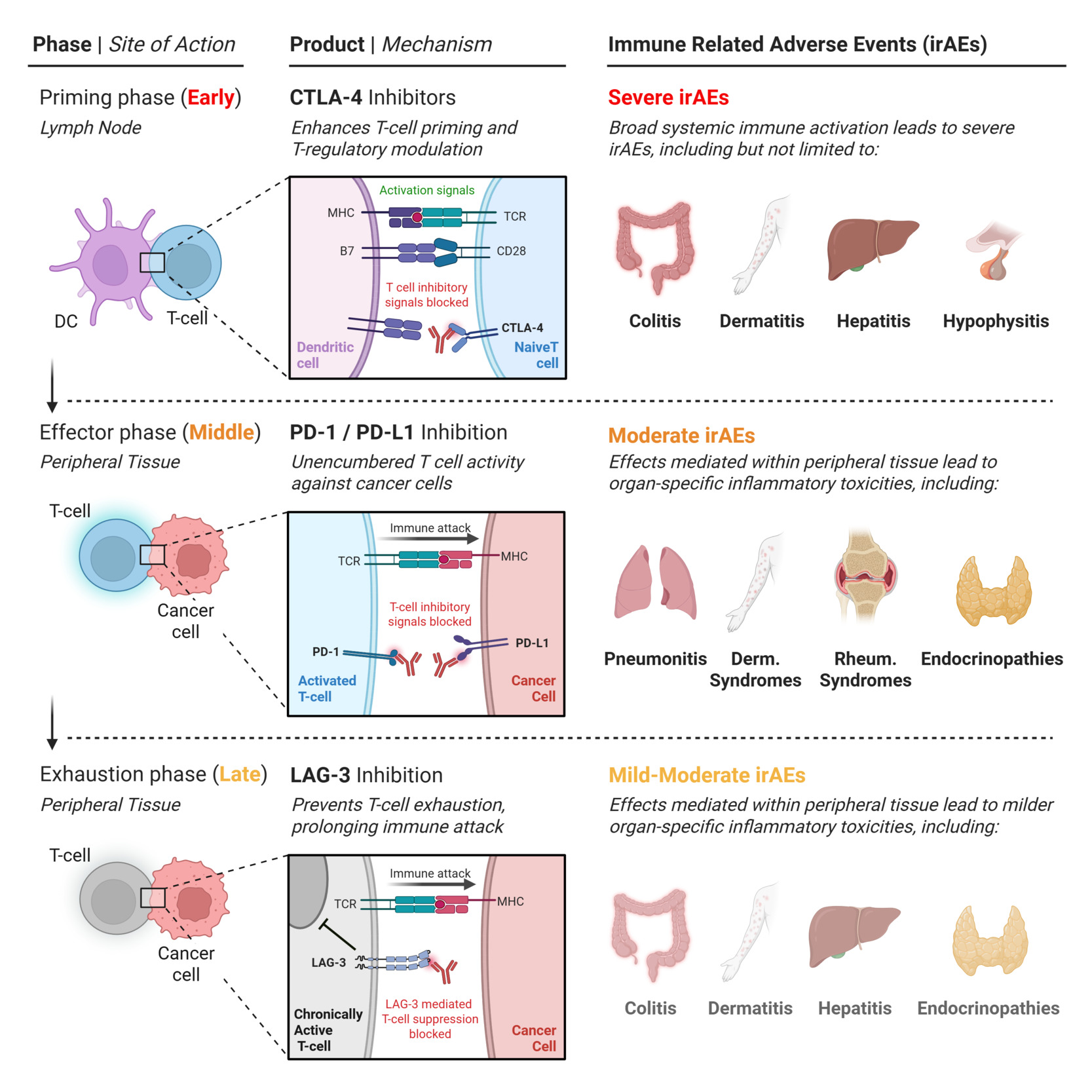
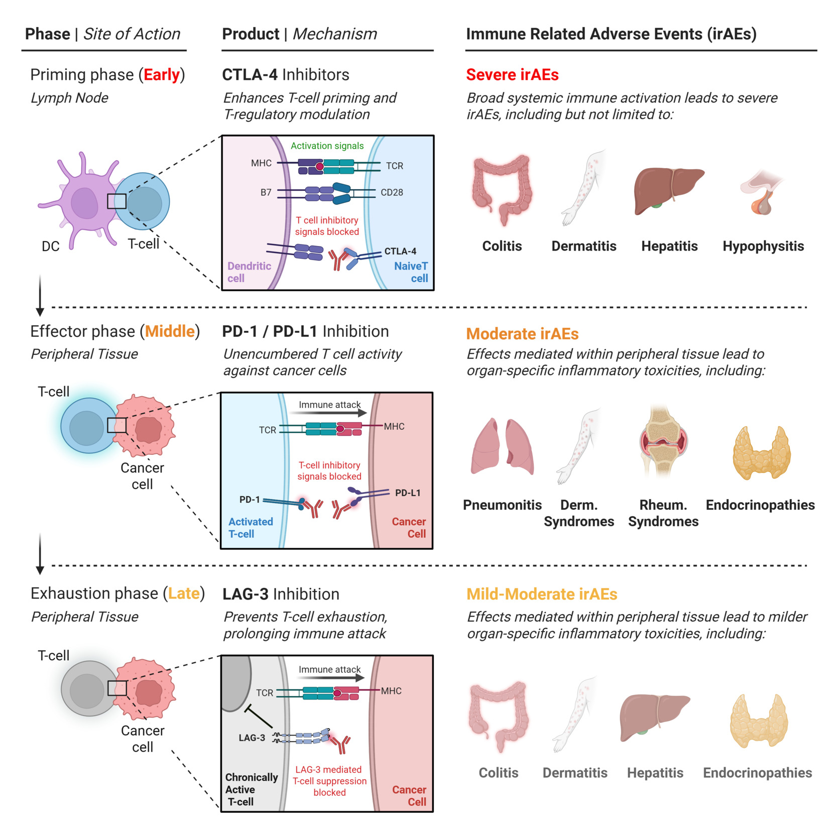
Immune checkpoint pathways regulate antitumor immunity at distinct stages of the T-cell response. CTLA-4 functions as an upstream regulator of early T-cell priming within secondary lymphoid organs.33–36 Effective activation requires antigen presentation by peptide–major histocompatibility complex (MHC) molecules together with CD28-mediated costimulation via CD80/CD86 on antigen-presenting cells. CTLA-4 competes with CD28 for CD80/CD86 with higher affinity, attenuating costimulatory signaling and limiting T-cell activation. It also mediates trans-endocytosis of CD80/CD86 from antigen-presenting cells, further suppressing antigen presentation.33,34 Pharmacologic CTLA-4 blockade disrupts this central tolerance checkpoint and produces broad systemic immune activation. Because CTLA-4 regulates the earliest stage of T-cell priming, its inhibition is associated with earlier onset and higher rates of immune-related adverse events (irAEs) than more distal checkpoints such as PD-1 or LAG-3.33,35–37 Clinically, CTLA-4 inhibitors are strongly associated with immune-mediated colitis and dermatitis, as well as hepatitis and hypophysitis, the latter often resulting in permanent endocrine dysfunction requiring long-term hormone replacement.15,38,39
PD-1/PD-L1 and Peripheral Tissue Tolerance
The PD-1 pathway regulates activated effector T cells within peripheral tissues and the tumor microenvironment (TME). Tumors frequently exploit this pathway through adaptive immune resistance. Inflammatory cytokines—particularly interferon-γ produced by activated CD8+ and CD4+ T cells and natural killer (NK) cells—induce PD-L1 expression on tumor and stromal cells. Engagement of PD-L1 with PD-1 on cytotoxic T cells suppresses T-cell receptor signaling and downstream effector function, limiting antitumor immunity.33,40,41 Therapeutic PD-1/PD-L1 blockade restores effector T-cell function at sites of antigen engagement while simultaneously disrupting PD-1-mediated peripheral tolerance in non-lymphoid tissues, predisposing to immune-related toxicities.15,33,40 Consequently, PD-1 inhibitors more commonly produce organ-specific inflammatory toxicities including endocrinopathies, pneumonitis, dermatologic inflammation, and rheumatologic syndromes.11,15,35 Pneumonitis is a particularly important complication and often occurs in tissues with preexisting injury such as prior thoracic irradiation or chronic lung disease.15,42,43
LAG-3 and T-Cell Exhaustion
During chronic antigen exposure, lymphocyte activation gene-3 (LAG-3) contributes to maintenance of T-cell exhaustion through redundant inhibitory signaling networks. Structurally homologous to the CD4 co-receptor, LAG-3 binds major histocompatibility complex (MHC) class II molecules with higher affinity than CD4, attenuating immunologic synapse formation and downstream T-cell receptor signaling.6,44,45 Within the TME, exhaustion is reinforced by additional ligands—including fibrinogen-like protein 1 (FGL1), galectin-3, and LSECtin—which suppress T-cell proliferation and cytokine secretion.46–48 Therapeutic LAG-3 blockade disrupts these inhibitory interactions and restores T-cell activity.5,6 When combined with PD-1 inhibition, LAG-3 blockade enhances T-cell reinvigoration while demonstrating lower rates of severe irAEs than CTLA-4-containing regimens.5,6,34–37,49,50
Determinants of Class-Specific Toxicity
Immune-mediated toxicities reflect the stages at which immune checkpoint therapy regulates T-cell activation. Their biologic positioning influences therapeutic synergy and the characteristic toxicity profiles observed across immunotherapy platforms.
Checkpoint Hierarchy, Toxicity Paradigms, and Checkpoint Layering
The biologic location of checkpoint pathways within the T-cell activation cascade provides a conceptual framework for understanding both therapeutic efficacy and immune-mediated toxicity. CTLA-4, PD-1, and LAG-3 operate at distinct stages of immune regulation, resulting in characteristic toxicity patterns and differing interactions when combined therapeutically. Immune checkpoints regulate antitumor immunity across the T-cell life cycle, forming a hierarchical network that maintains immune tolerance while permitting effective immune activation.33–37,49 CTLA-4 restrains early T-cell priming within lymphoid organs, PD-1 limits effector activity in peripheral tissues, and LAG-3 reinforces exhaustion in chronically stimulated T-cell populations within the TME.33,34,40,44–48 Accordingly, checkpoint blockades produce distinct toxicity patterns. CTLA-4 inhibition induces broad systemic immune activation and the highest rates of multisystem irAEs, whereas PD-1 inhibition more commonly causes organ-specific inflammatory toxicities reflecting loss of peripheral immune tolerance. LAG-3-containing regimens generally demonstrate a comparatively moderate toxicity profile consistent with localized modulation of exhausted effector T cells.5,6,15,35–37,49,50
Divergent Toxicity Paradigms: irAEs versus Immune Effector Cytokine Syndromes
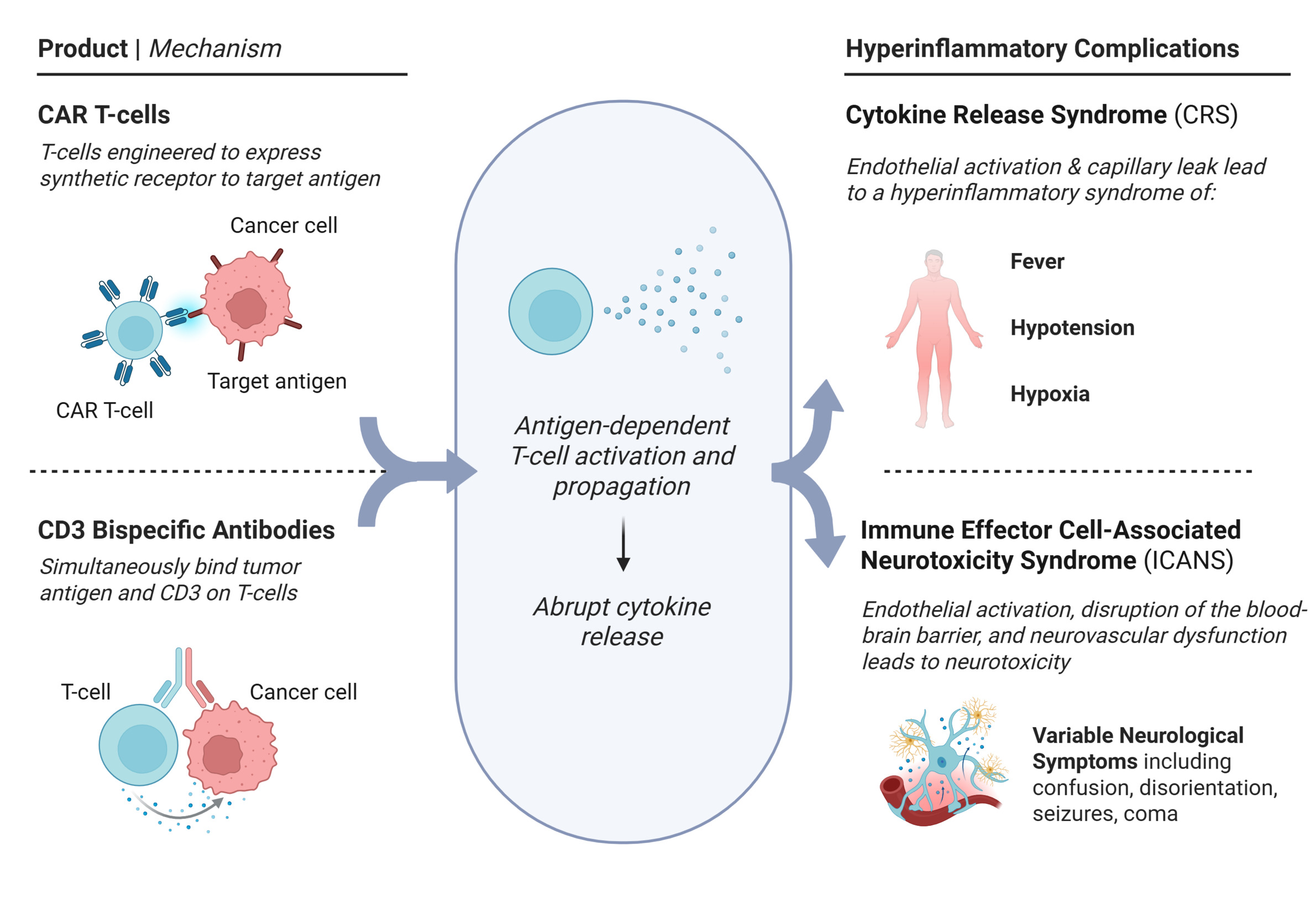
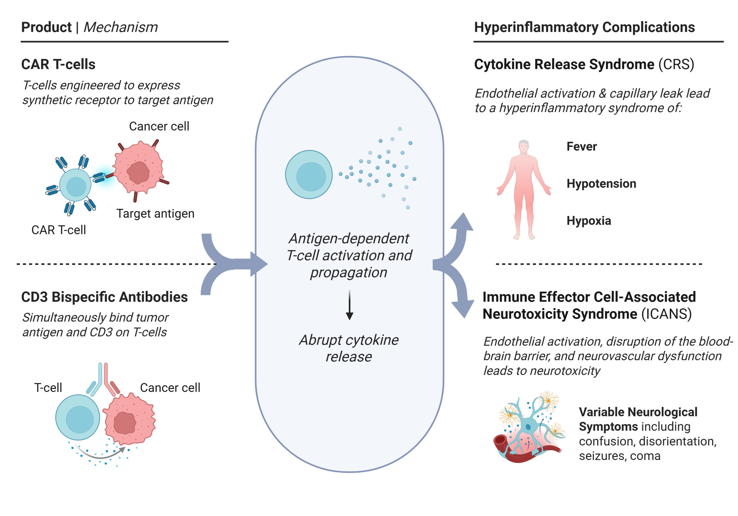
A key clinical distinction exists between autoimmune-like irAEs induced by ICIs and the acute toxicities associated with IEC therapies such as CAR T-cell therapy and CD3-engaging bispecific T-cell engagers. ICIs gradually erode peripheral immune tolerance, unmasking autoreactive immune responses across multiple tissues. By contrast, IEC therapies trigger rapid antigen-dependent T-cell expansion with abrupt cytokine release—particularly IL-6, IFN-γ, and TNF-α.14,18,33 This cytokine surge drives endothelial activation, capillary leak, and neurovascular dysfunction, manifesting clinically as CRS and ICANS.14,18,51 Checkpoint-associated irAEs are therefore managed primarily with immunosuppression aimed at restoring immune tolerance, whereas CRS and ICANS represent states of immune hyperactivation treated mainly with cytokine-directed therapies—most commonly IL-6 blockade—and intensive supportive care.14,18,33,51
Checkpoint Layering: Complementarity versus Redundancy
The clinical impact of checkpoint combinations depends on whether targeted pathways are biologically complementary or functionally redundant. Dual CTLA-4/PD-1 blockade enhances antitumor immunity by coupling expanded T-cell priming with sustained peripheral effector activity and improves long-term outcomes in melanoma, albeit with substantially increased rates of high-grade irAEs.33,36,37,52 In contrast, PD-1/LAG-3 inhibition targets exhausted effector T-cell populations within the TME and provides incremental clinical benefit with an intermediate toxicity profile while avoiding the severe immune toxicity characteristic of CTLA-4-containing regimens.5,6,36,37,49 TIGIT inhibition—a next-generation checkpoint strategy with functional overlap with PD-(L)1 signaling—has produced inconsistent benefit in late-phase trials when combined with PD-L1 blockade. These findings highlight the challenges of targeting partially redundant inhibitory pathways where enhanced immune activation may not translate into improved survival outcomes.53–55
The types of immunotherapies and their contrasting toxicity paradigms are summarized in Figures 1-3.
Organ-Specific Immune-Related Adverse Events Associated with Immune Checkpoint Inhibition
The immunologic mechanisms of checkpoint inhibition described above translate clinically into a broad spectrum of organ-specific irAEs. Unlike toxicities caused by cytotoxic chemotherapy, these complications arise from systemic immune disinhibition rather than direct tissue injury. irAEs vary widely in timing, severity, reversibility, and clinical consequences across organ systems. Although many events are low grade and manageable, others carry risks of irreversible organ dysfunction or death. Effective management therefore depends on early recognition, phenotype-directed evaluation, timely escalation of immunosuppression, and coordinated multidisciplinary care.11,15
Dermatologic Toxicity
Cutaneous irAEs are the most common toxicities of ICI therapy, occurring in approximately 30–50% of patients. They typically arise early, most often within the first 2–8 weeks of treatment, although delayed presentations—including events after treatment discontinuation—have been reported, reflecting durable immune modulation induced by checkpoint blockade.56–58 The clinical spectrum includes inflammatory and autoimmune phenotypes. Common inflammatory eruptions such as maculopapular rash, lichenoid dermatitis, and psoriasis-like lesions are largely mediated by cytotoxic T-cell–driven keratinocyte injury. Autoimmune manifestations—including vitiligo-like depigmentation, alopecia areata, and bullous pemphigoid—reflect melanocyte-directed inflammation or pathogenic autoantibody production.56–58 Less frequent but clinically important entities include cutaneous vasculitis, subacute cutaneous lupus erythematosus, and dermatomyositis-like syndromes.57 Vitiligo-like depigmentation in melanoma is strongly associated with favorable oncologic outcomes and generally requires no intervention beyond reassurance and supportive skin care.57 More broadly, development of cutaneous irAEs has been correlated with improved survival across multiple tumor types, supporting their role as markers of effective immune activation rather than treatment failure.15,16,57,59
Management is guided primarily by body surface area (BSA) involvement and functional impact. Limited eruptions involving <10% BSA without blistering or mucosal involvement (grade 1) are typically managed with topical corticosteroids, emollients, and antihistamines without interruption of immunotherapy.13,51,60,61 Grade 2 toxicity involving 10–30% BSA or functional limitation usually requires temporary immunotherapy interruption and systemic corticosteroids (prednisone 0.5–1 mg/kg/day) with gradual tapering after improvement.13,51,60,61 Severe dermatologic irAEs (>30% BSA, blistering, or mucosal ulceration; grade 3–4) require hospitalization, dermatology consultation, and intravenous methylprednisolone 1–2 mg/kg/day.13,62 Early recognition of severe cutaneous adverse reactions such as Stevens–Johnson syndrome, toxic epidermal necrolysis, and DRESS is critical because these conditions require permanent discontinuation of ICIs and urgent multidisciplinary management.61–63
Pruritus is common and may be disproportionately symptomatic. Because immune-mediated pruritus is often cytokine-driven rather than histaminergic, antihistamines may be insufficient.57 Neuropathic agents such as gabapentin or pregabalin may provide benefit, while steroid-refractory cases may respond to biologic therapies such as dupilumab or omalizumab.64–66 Autoimmune blistering disorders—particularly bullous pemphigoid—often require escalation beyond corticosteroids with rituximab or intravenous immunoglobulin to suppress pathogenic autoantibody production.62,67
Rechallenge decisions depend on phenotype and severity. Patients with uncomplicated inflammatory eruptions that fully resolve may be cautiously rechallenged after corticosteroid tapering, whereas rechallenge is contraindicated following severe cutaneous adverse reactions and generally avoided after severe autoimmune blistering disease. Long-term sequelae such as xerosis, pigmentary change, nail dystrophy, and scarring alopecia support ongoing dermatologic follow-up during survivorship.13,57,63
Pulmonary Toxicity
ICI-associated pneumonitis is an uncommon but potentially life-threatening irAE and a leading cause of immunotherapy-related mortality. It occurs in approximately 2–5% of patients receiving ICI monotherapy, with higher incidence in combination regimens. Risk increases in patients with pre-existing interstitial lung disease, prior thoracic radiation, or significant smoking history.7–15,42,43 The pathogenesis reflects dysregulated T-cell activation and cytokine-mediated alveolar inflammation resulting in impaired gas exchange.15,43 High-resolution CT most commonly demonstrates an organizing pneumonia pattern characterized by peripheral ground-glass opacities and consolidations with lower-lobe predominance. Other patterns include nonspecific interstitial pneumonia, hypersensitivity pneumonitis–like disease, and diffuse alveolar damage, the latter associated with severe respiratory failure and high mortality.42,43,68Diagnosis requires exclusion of infection, pulmonary embolism, radiation injury, and tumor progression. High-resolution CT is central to evaluation, and microbiologic testing is essential in symptomatic patients. Bronchoscopy with bronchoalveolar lavage is frequently performed to exclude opportunistic pathogens, although lymphocytic predominance alone is not diagnostic.13,43
Grading is based primarily on clinical severity, particularly symptoms and oxygen requirement.11,13 In addition, ESMO guidelines incorporate radiographic extent of lung involvement as a supportive criterion, with <25% lung involvement generally corresponding to grade 1–2 disease and ≥25–50% or more extensive bilateral involvement associated with grade ≥3 severity.62 Grade 1 pneumonitis typically presents as asymptomatic radiographic abnormalities and may be managed with surveillance and temporary immunotherapy interruption. Grade 2 pneumonitis requires withholding immunotherapy and initiating systemic corticosteroids (prednisone 1–2 mg/kg/day) with reassessment within 48–72 hours. Grade 3 pneumonitis requires hospitalization, oxygen support, permanent immunotherapy discontinuation, and intravenous methylprednisolone 1–2 mg/kg/day. Grade 4 disease represents life-threatening respiratory failure requiring intensive care management.11,13,61
Steroid-refractory pneumonitis warrants escalation of immunosuppression. Mycophenolate mofetil is commonly used once infection has been excluded, while IVIG, tocilizumab, or cyclophosphamide may be considered in refractory cases. Infliximab is generally avoided because of infection risk.13,62,69,70 Rechallenge with ICIs may be considered after complete resolution of grade 1 pneumonitis and in selected cases of resolved grade 2 disease but is contraindicated following grade 3–4 toxicity due to high recurrence risk.11,13
Clinical Pearl: Pneumonitis remains one of the leading causes of fatal irAEs. Because radiographic findings are often nonspecific, exclusion of infection and tumor progression is critical before escalating immunosuppression. Early recognition and treatment may prevent progression to respiratory failure.
Gastrointestinal Toxicity
Immune-mediated gastrointestinal toxicity is one of the most clinically significant irAEs and occurs most frequently with CTLA-4–containing regimens and combination checkpoint blockade. Although median onset typically occurs within 4–8 weeks of therapy initiation, delayed presentations—including events after prolonged exposure or treatment discontinuation—are increasingly recognized, reflecting sustained immune activation induced by checkpoint inhibition.15,42,62 At the tissue level, immune-mediated colitis reflects T-cell–driven epithelial injury and disruption of mucosal immune homeostasis. Histologic features frequently resemble inflammatory bowel disease, including cryptitis, crypt abscesses, and lymphocytic infiltration of the lamina propria.55,71 Mechanistic studies suggest that CTLA-4 blockade disrupts intestinal immune tolerance through depletion of regulatory T cells and enhanced effector T-cell activation within the gut mucosa, providing a biologic explanation for the higher incidence of colitis with CTLA-4–based regimens. Clinically, toxicity severity is defined using CTCAE criteria, with diarrhea graded by stool frequency above baseline and colitis incorporating features such as abdominal pain, hematochezia, mucus in stool, ileus, or peritoneal signs.11,72
Mild diarrhea—typically fewer than four stools per day above baseline (grade 1)—is often self-limited and managed conservatively with hydration, dietary modification, and antimotility agents while continuing immunotherapy with close monitoring.11,62 Increasing stool frequency to four to six stools per day above baseline or development of abdominal symptoms (grade 2) suggests clinically meaningful intestinal inflammation and should prompt further evaluation. Diagnostic assessment requires exclusion of infectious etiologies, particularly Clostridioides difficile and other enteric pathogens that may mimic immune-mediated colitis in immunocompromised patients.11,62,73 Biomarkers such as fecal calprotectin correlate with inflammatory activity and endoscopic severity, and endoscopic evaluation with mucosal biopsy is frequently pursued when symptoms persist or diagnosis remains uncertain.62,73,74
When immune-mediated colitis is suspected, temporary interruption of immunotherapy and systemic corticosteroids are generally required. Oral prednisone 1–2 mg/kg/day is commonly used for moderate disease, whereas patients with more severe presentations—typically ≥7 stools per day above baseline, incontinence, or hospitalization (grade 3)—require inpatient management and intravenous methylprednisolone.11,61,62 Clinical improvement is generally expected within 48–72 hours; lack of response suggests steroid-refractory disease. Contemporary guidelines increasingly recommend early escalation to biologic therapy for steroid-refractory immune-mediated colitis, most commonly infliximab or vedolizumab, to prevent disease progression and reduce prolonged corticosteroid exposure.11,12,62,74 Clinical reassessment is recommended within 24–48 hours after initiation of high-dose corticosteroids, and lack of meaningful improvement—defined by persistent diarrhea, ongoing abdominal pain, or failure of stool frequency to decline—should prompt early initiation of biologic therapy rather than prolonged steroid monotherapy.11,62,74
Infliximab, a TNF-α inhibitor, and vedolizumab, a gut-selective α4β7 integrin antagonist, both demonstrate efficacy in steroid-refractory disease. Infliximab is often preferred for rapid disease control, whereas vedolizumab may be favored in patients at increased risk of systemic infection due to its gut-selective immunologic effect.11,62,74 Early biologic escalation—typically within 48–72 hours of inadequate steroid response—has been associated with improved symptom control, reduced hospitalization duration, and decreased cumulative corticosteroid exposure.12,62 Rare but life-threatening complications—including bowel perforation, toxic megacolon, hemorrhage, or sepsis (grade 4 colitis)—require urgent multidisciplinary management and permanent discontinuation of checkpoint therapy.11,62
Incomplete response despite biologic therapy warrants reassessment, including evaluation for cytomegalovirus colitis, which may complicate immunosuppression and requires biopsy confirmation and antiviral therapy.74,75In highly refractory cases, fecal microbiota transplantation has demonstrated benefit in selected patients by restoring intestinal microbial homeostasis and modulating mucosal immune responses.76,77 Rechallenge with checkpoint inhibition may be cautiously considered after complete resolution of moderate colitis, although recurrence remains common—particularly following CTLA-4 re-exposure—and retreatment is generally avoided after severe or life-threatening gastrointestinal toxicity.11,74
Clinical Pearl: Contemporary management has shifted toward earlier biologic escalation for steroid-refractory colitis. In patients without meaningful improvement within 48–72 hours of initiating corticosteroids, infliximab or vedolizumab should be considered rather than prolonged corticosteroid monotherapy, which may increase the risk of infectious complications and cumulative steroid exposure.
Endocrine Toxicity
Endocrine irAEs represent a distinct category of ICI toxicity characterized by insidious onset, frequent irreversibility, and long-term management implications rather than acute inflammatory morbidity. Incidence varies by regimen, occurring in approximately 5–15% of patients receiving PD-1/PD-L1 monotherapy and rising to nearly 30% with combination regimens incorporating CTLA-4 inhibition. Unlike many other irAEs, endocrine toxicities often reflect permanent destruction of hormone-producing cells, requiring lifelong physiologic hormone replacement and survivorship-focused care.15,39,62 The pathogenesis involves dysregulated T-cell infiltration and cytokine-mediated glandular injury, with a characteristic association between checkpoint class and phenotype: CTLA-4 blockade is most strongly linked to lymphocytic hypophysitis, whereas PD-1/PD-L1 inhibition more commonly induces destructive thyroiditis and insulin-deficient diabetes mellitus. Thyroid dysfunction is the most frequent manifestation, while hypophysitis and adrenal insufficiency remain clinically critical because of their potential to cause life-threatening metabolic decompensation if unrecognized.15,39,62
Clinical recognition can be challenging because presenting symptoms—fatigue, anorexia, nausea, dizziness, hypotension, weight change, or mood disturbance—are nonspecific and often attributed to malignancy or systemic therapy. Consequently, structured biochemical surveillance is essential. Baseline and periodic evaluation of thyroid-stimulating hormone (TSH), free thyroxine (T4), morning cortisol, and glucose is recommended during checkpoint therapy.11,61,62 When adrenal insufficiency is suspected, measurement of morning cortisol and adrenocorticotropic hormone (ACTH) helps distinguish primary from secondary etiologies. In suspected hypophysitis, comprehensive pituitary hormone evaluation is required; pituitary MRI may reveal gland enlargement or stalk thickening, although normal imaging does not exclude clinically significant disease.39,62 Management focuses on physiologic hormone replacement rather than prolonged immunosuppression. Immune-mediated thyroiditis often follows a biphasic course, beginning with transient thyrotoxicosis managed symptomatically with beta-blockade and progressing to permanent hypothyroidism requiring levothyroxine replacement titrated to biochemical targets.15,62 A critical principle is prioritizing glucocorticoid replacement when adrenal insufficiency is suspected, particularly in central hypothyroidism, because levothyroxine initiation before cortisol repletion may precipitate adrenal crisis.39,62
Hypophysitis and adrenal insufficiency typically require lifelong glucocorticoid replacement, most commonly hydrocortisone 15–25 mg/day in divided doses, with patient education regarding stress dosing and emergency parenteral steroid administration during major illness or procedures.39,62 ICI-associated diabetes mellitus, though less common, often presents abruptly with diabetic ketoacidosis and reflects irreversible β-cell destruction; management therefore requires lifelong insulin therapy, and corticosteroids do not reverse established endocrine injury.62,78 Unlike pulmonary, neurologic, or cardiac irAEs, endocrine toxicities rarely necessitate permanent discontinuation of immune checkpoint therapy once stabilized. Rechallenge is generally feasible when hormonal deficits are adequately controlled, emphasizing recognition of endocrine irAEs as chronic endocrine comorbidities integrated into survivorship care rather than treatment-limiting complications.11,62
Hepatic Toxicity
Hepatic toxicity associated with ICIs most commonly manifests as immune-mediated hepatitis and represents an important cause of treatment interruption or discontinuation. Incidence is approximately 2–5% with PD-1/PD-L1 monotherapy and increases to 10–20% with combination checkpoint blockade, particularly regimens incorporating CTLA-4 inhibition. Hepatitis typically develops within the first 6–12 weeks of therapy, although delayed presentations—including events after treatment discontinuation—are increasingly recognized. Risk appears higher in patients receiving combination immunotherapy and in those with underlying liver disease, hepatic metastases, or prior hepatotoxic treatments.15,62,79 The pathogenesis reflects immune-mediated hepatocellular injury driven by enhanced T-cell activation, loss of immune tolerance, and cytokine-mediated inflammation. Histopathology most commonly demonstrates panlobular hepatitis with lymphocytic infiltration, lobular necrosis, and portal inflammation, resembling autoimmune hepatitis but typically lacking classical autoantibody profiles. Cholestatic and mixed hepatocellular–cholestatic patterns may also occur, reflecting heterogeneous immune mechanisms.79
ICI-associated hepatitis is frequently asymptomatic and detected through routine laboratory monitoring. When present, symptoms may include fatigue, anorexia, right upper quadrant discomfort, nausea, pruritus, jaundice, or dark urine, while severe disease can progress to coagulopathy, hepatic encephalopathy, or acute liver failure. Cholestatic presentations often follow a more protracted course and may demonstrate delayed biochemical recovery.62,79 Evaluation requires systematic exclusion of alternative etiologies. Recommended assessment includes viral serologies (hepatitis A, B, C, and E; cytomegalovirus; Epstein–Barr virus), autoimmune markers (antinuclear antibody, anti–smooth muscle antibody, immunoglobulin G), medication and alcohol history, and metabolic testing when appropriate. Abdominal ultrasonography or cross-sectional imaging is performed to exclude biliary obstruction, vascular abnormalities, or tumor progression. Liver biopsy is not routinely required but may be considered in cases of diagnostic uncertainty, atypical biochemical patterns, or inadequate response to corticosteroids.11,62
Severity is graded primarily according to serum aminotransferase and bilirubin levels. Grade 1 hepatitis is defined by AST or ALT >1 to ≤3 × the upper limit of normal (ULN) with bilirubin ≤1.5 × ULN. Grade 2 disease involves AST or ALT >3 to ≤5 × ULN and/or bilirubin >1.5 to ≤3 × ULN. Grade 3 hepatitis is characterized by AST or ALT >5 to ≤20 × ULN or bilirubin >3 to ≤10 × ULN, often accompanied by symptoms, whereas grade 4 toxicity reflects life-threatening hepatic injury with AST or ALT >20 × ULN, bilirubin >10 × ULN, or evidence of hepatic failure including coagulopathy or encephalopathy.11,62
Routine monitoring of liver function tests—including AST, ALT, alkaline phosphatase, bilirubin, and international normalized ratio—is essential during immunotherapy. Rapid enzyme escalation, rising bilirubin, or coagulopathy requires urgent evaluation. Management is guided by biochemical severity and clinical trajectory. Grade 1 hepatitis is generally managed with continued immunotherapy and close laboratory surveillance. Grade 2 disease requires temporary interruption of ICI therapy and initiation of oral corticosteroids (prednisone 0.5–1 mg/kg/day) with laboratory reassessment every 3–5 days. Grade 3–4 hepatitis necessitates permanent discontinuation of immunotherapy and high-dose corticosteroids, typically intravenous methylprednisolone 1–2 mg/kg/day, often with hospitalization and monitoring for hepatic decompensation. Corticosteroids should be tapered gradually over approximately 4–6 weeks after biochemical improvement to reduce relapse risk.11,61,62
Steroid-refractory hepatitis, defined by lack of improvement after 3–5 days of high-dose corticosteroids, warrants escalation of immunosuppression. Mycophenolate mofetil (500–1000 mg twice daily) is the preferred second-line therapy and has demonstrated efficacy in retrospective studies. TNF inhibitors such as infliximab are generally avoided because of potential hepatotoxicity. Alternative agents—including tacrolimus or azathioprine—may be considered in selected refractory cases in collaboration with hepatology specialists.11,62,79 Although many patients achieve biochemical resolution, a subset develops persistent liver injury including chronic cholestasis, portal inflammation, or fibrosis, requiring ongoing follow-up. Early hepatology involvement is recommended in cases of high-grade toxicity, steroid-refractory hepatitis, or underlying chronic liver disease.62,79
Rechallenge following immune-mediated hepatitis remains controversial. Retrospective studies suggest recurrence rates of approximately 25–40%, with higher risk after grade ≥3 toxicity. Rechallenge may be cautiously considered in selected patients with prior grade 2 hepatitis who achieve complete biochemical recovery and lack effective alternative therapies. However, rechallenge is generally discouraged after grade 3–4 hepatitis, particularly in patients with prior steroid-refractory disease or persistent hepatic dysfunction.62,80
Cardiovascular Toxicity
Immune-related cardiovascular toxicities are uncommon but among the most feared irAEs because of their high morbidity and mortality. Myocarditis is the prototypical manifestation and typically presents early, often within the first 4–6 weeks after initiation of ICIs. Risk is markedly increased with combination checkpoint blockade, reflecting broader immune activation and loss of peripheral tolerance.81–83 Clinical presentation is heterogeneous and may initially be subtle. Patients may report fatigue, dyspnea, chest discomfort, or palpitations, while others present abruptly with malignant ventricular arrhythmias, high-grade atrioventricular block, cardiogenic shock, or sudden cardiac death.81,82 A particularly high-risk phenotype is concurrent myocarditis, myositis, and immune-related myasthenia gravis (“triple-M overlap”), which carries a poor prognosis and requires urgent multidisciplinary management.84
Diagnosis requires a high index of suspicion because early ECG and echocardiographic findings may be nonspecific or normal. Evaluation includes serial high-sensitivity troponin and natriuretic peptides, electrocardiography, transthoracic echocardiography, and cardiac MRI with late gadolinium enhancement and parametric mapping when feasible. Although endomyocardial biopsy remains the diagnostic gold standard, treatment should not be delayed when myocarditis is strongly suspected.81,83 Management constitutes a medical emergency. ICIs should be permanently discontinued and high-dose systemic corticosteroids initiated promptly. Pulse intravenous methylprednisolone (commonly 1000 mg daily for 3 days) is frequently used, followed by high-dose oral or intravenous corticosteroids (1–2 mg/kg/day) with gradual tapering guided by biomarker trends and clinical stability.80,82 Early steroid initiation is associated with improved outcomes and fewer arrhythmic complications.81,85
Patients without rapid biochemical or clinical improvement within 24–48 hours are considered steroid-refractory and require escalation of immunosuppression. Abatacept has emerged as a rational second-line therapy targeting T-cell costimulation and has demonstrated promising activity in refractory cases. Additional options include IVIG, mycophenolate mofetil, or antithymocyte globulin in selected patients. Concurrent management of heart failure, arrhythmias, and conduction abnormalities—including temporary pacing or mechanical circulatory support—may be required.82,83Given the risk of recurrence and catastrophic outcomes, rechallenge with ICIs after myocarditis is strongly discouraged regardless of initial severity. Long-term cardiology follow-up is recommended, as residual ventricular dysfunction, arrhythmias, and delayed cardiomyopathy may persist despite immunologic control.81,82
Clinical Pearl: ICI myocarditis may present with preserved or only mildly reduced left ventricular ejection fraction despite life-threatening disease. Clinicians should maintain a low threshold for troponin testing, cardiac MRI, and early pulse-dose corticosteroid therapy, as delayed treatment is associated with worse outcomes. Although abatacept has emerged as a promising option for steroid-refractory myocarditis, current evidence remains largely based on retrospective studies and case series.
Neurologic Toxicity
Neurologic irAEs are uncommon but among the most clinically complex and potentially severe toxicities associated with immune checkpoint inhibition. They most often occur within the first 6–12 weeks of therapy, although delayed presentations months after treatment initiation or even after discontinuation are increasingly recognized. Risk appears higher with combination checkpoint blockade and in patients who develop overlap syndromes involving skeletal muscle or cardiac inflammation.86,87The clinical spectrum involves both the central and peripheral nervous systems. Central manifestations include immune-mediated encephalitis, aseptic meningitis or meningoencephalitis, cerebellitis, transverse myelitis, demyelinating syndromes resembling multiple sclerosis relapse, and vasculitic processes. Peripheral involvement includes Guillain–Barré–like polyradiculoneuropathy, chronic inflammatory demyelinating polyneuropathy–like syndromes, painful sensory or small-fiber neuropathy, cranial neuropathies, autonomic dysfunction, and immune-related myasthenia gravis (irMG).88,89 Among these entities, irMG carries particularly high morbidity and mortality, especially when occurring with concurrent myositis or myocarditis. This overlap syndrome may progress rapidly to respiratory compromise and therefore represents a neurologic emergency requiring urgent multidisciplinary management.84
Diagnosis is syndrome-directed and requires exclusion of infection, metabolic derangements, and tumor progression. Evaluation typically includes comprehensive neurologic examination with assessment of bulbar and respiratory function, MRI of the brain and/or spine with contrast, and cerebrospinal fluid analysis. Electrodiagnostic testing is essential when neuropathic or neuromuscular junction disorders are suspected, while electroencephalography is useful in encephalopathy or seizure-predominant presentations. In suspected irMG, evaluation should include creatine kinase, troponin, ECG, myasthenia antibodies (AChR and MuSK), and serial respiratory mechanics, recognizing that antibody testing may be negative despite clinically significant disease.90–92
Management depends on clinical severity and requires early intervention to prevent irreversible neurologic injury. Mild sensory symptoms without functional limitation may be monitored with close follow-up. When symptoms interfere with instrumental activities of daily living—such as gait instability, moderate weakness, diplopia, or dysphagia—ICIs are typically withheld and systemic corticosteroids initiated (prednisone 0.5–1 mg/kg/day or equivalent).61,62,87 Severe presentations—including encephalitis, myelitis, rapidly progressive neuropathy, bulbar dysfunction, autonomic instability, seizures, or respiratory compromise—require hospitalization and high-dose intravenous corticosteroids (methylprednisolone 1–2 mg/kg/day). Pulse therapy (1000 mg/day for 3–5 days) is often used in fulminant encephalitis or transverse myelitis, followed by gradual tapering over 6–12 weeks to reduce relapse risk.62,87
Clinical reassessment should occur within 24–48 hours after initiation of high-dose corticosteroids, and lack of objective improvement—particularly persistent weakness, bulbar symptoms, or evolving respiratory compromise—should prompt immediate escalation to adjunctive immunotherapy rather than prolonged steroid monotherapy. IVIG or plasmapheresis should be initiated promptly (typically within 24–48 hours of inadequate steroid response) in suspected irMG or Guillain–Barré–like syndromes, especially when bulbar dysfunction or respiratory involvement is present. Early use of these therapies is critical, as neurologic irAEs may progress more rapidly than other immune toxicities and are associated with higher morbidity and mortality.62,84Refractory cases may require additional immunosuppressive agents such as mycophenolate mofetil, rituximab, or cyclophosphamide, although evidence is limited and management should be individualized. Close neurologic monitoring with a low threshold for ICU-level care is essential, particularly for patients with irMG, where rapid progression to respiratory failure can occur. Serial assessment of respiratory function is recommended.12,62 Importantly, medications that impair neuromuscular junction function (e.g., fluoroquinolones, aminoglycosides, magnesium, beta-blockers) should be avoided, as they may precipitate or worsen neuromuscular weakness.62,84
Given the risk of recurrence and severe outcomes, rechallenge with ICIs is generally discouraged after severe neurologic irAEs, particularly encephalitis, myelitis, Guillain–Barré–like syndromes, or irMG. Carefully selected patients with mild, fully resolved sensory neuropathy may be considered for rechallenge following multidisciplinary risk–benefit assessment and close neurologic monitoring.11,62
Clinical Pearl: Neurologic irAEs often progress more rapidly than other immune toxicities. In suspected immune-related myasthenia gravis, Guillain–Barré–like syndrome, or overlap syndromes, early IVIG or plasmapheresis should not be delayed while awaiting a response to corticosteroids.
Rheumatologic and Musculoskeletal Toxicity
Rheumatologic and musculoskeletal irAEs are among the most common chronic toxicities of immune checkpoint inhibition and represent an important source of long-term morbidity. They typically emerge within the first 2–4 months of therapy but may occur at any time during treatment or after discontinuation. Their true incidence is likely underestimated because symptoms frequently overlap with cancer-related fatigue, degenerative joint disease, or paraneoplastic syndromes.93The clinical spectrum is broad and includes inflammatory arthritis, polymyalgia rheumatica (PMR)–like syndromes, seronegative rheumatoid arthritis–like disease, spondyloarthritis-like phenotypes, tenosynovitis, sicca syndrome, sarcoid-like granulomatous disease, systemic vasculitis, and inflammatory myopathies. Inflammatory arthritis and PMR-like syndromes are most frequent and often persist despite discontinuation of immunotherapy, reflecting durable immune dysregulation. Myositis, although less common, is clinically significant—particularly when occurring with myocarditis or immune-related myasthenia gravis.94,95
Evaluation requires exclusion of metastatic disease, infection, crystal arthropathy, and degenerative conditions. Assessment typically includes inflammatory markers (ESR, CRP), creatine kinase measurement when myositis is suspected, and selective autoantibody testing (ANA, RF, anti-CCP), recognizing that many patients remain seronegative. Ultrasound or MRI may help confirm synovitis, tenosynovitis, or myositis when diagnosis is uncertain, and early rheumatology consultation is recommended for persistent or moderate-to-severe disease.94,96 Management is guided by symptom severity and functional impairment. Mild arthralgia or myalgia without functional limitation may be treated with NSAIDs, acetaminophen, and physical therapy while continuing immunotherapy. When symptoms limit instrumental activities of daily living, ICIs are typically withheld and low-to-moderate dose corticosteroids initiated (prednisone 10–20 mg/day) with gradual tapering as symptoms improve.11,13,61,93 Severe inflammatory arthritis, vasculitis, or myositis generally requires higher-dose corticosteroids (0.5–1 mg/kg/day), with hospitalization when systemic involvement or overlap syndromes are present.11,13,94
Because many rheumatologic irAEs follow a chronic or relapsing course, steroid-sparing therapy is often required. Conventional DMARDs, including methotrexate, sulfasalazine, and hydroxychloroquine, are commonly used for persistent inflammatory arthritis or PMR-like syndromes. Refractory disease may require biologic therapy, such as TNF inhibitors (e.g., infliximab) or IL-6 receptor antagonists (e.g., tocilizumab), in coordination with oncology and rheumatology specialists. Rechallenge with ICIs may be considered in selected patients whose symptoms are controlled with low-dose corticosteroids or stable DMARD therapy, but is generally avoided after severe myositis, systemic vasculitis, or overlap syndromes.93,94,96,97
Renal Toxicity
Immune-mediated renal toxicity associated with ICIs most commonly manifests as acute interstitial nephritis, although glomerulonephritis, vasculitis, and thrombotic microangiopathy have also been reported. Presentation is often insidious, with asymptomatic serum creatinine elevation detected during routine monitoring, frequently accompanied by sterile pyuria and mild proteinuria.12,61,62,98–100Evaluation aims to confirm immune-mediated injury while excluding alternative causes of renal dysfunction. Serial creatinine monitoring and urinalysis with microscopy are essential once renal dysfunction is detected, as inflammatory findings may support interstitial nephritis and help exclude infection or obstruction. Renal imaging is generally reserved for suspected structural disease, while kidney biopsy should be considered in cases of diagnostic uncertainty, atypical urinary findings, heavy proteinuria, or severe or rapidly progressive renal dysfunction.11–13,61,62,98–100
Baseline urinalysis before ICI initiation is not routinely recommended because of limited predictive value and the potential for nonspecific abnormalities. Accordingly, urinalysis is typically obtained after creatinine elevation to guide diagnostic evaluation and management.11–13,62,101 When serum creatinine rises above the upper limit of normal but remains ≤1.5 times baseline, renal dysfunction may be managed with continued immunotherapy, close monitoring, and removal of potential nephrotoxic medications such as proton pump inhibitors, NSAIDs, iodinated contrast, and certain antibiotics.11–13,62,101 Creatinine elevation >1.5 times baseline generally warrants withholding immunotherapy and initiating systemic corticosteroids (prednisone 0.5–1 mg/kg/day).11–13,61,62 Severe injury—defined by creatinine >3 times baseline, absolute creatinine >4 mg/dL, rapidly progressive renal failure, or biopsy-proven severe nephritis—requires permanent ICI discontinuation, hospitalization, high-dose corticosteroids, and nephrology co-management, including renal replacement therapy when indicated.11–13,62,101 After renal recovery, corticosteroids should be tapered gradually over several weeks to reduce relapse risk. Steroid-refractory disease may require escalation to additional immunosuppressive therapy such as mycophenolate mofetil, and ICI rechallenge is individualized, typically considered only after complete renal recovery with close laboratory surveillance.11–13,62
Hematologic Toxicity
Hematologic irAEs are rare but potentially life-threatening and encompass a diverse group of immune-mediated cytopenias and marrow disorders. Reported entities include autoimmune hemolytic anemia (AIHA), immune thrombocytopenia (ITP), thrombotic microangiopathy (TMA), neutropenia, aplastic anemia, pure red cell aplasia, and hemophagocytic lymphohistiocytosis (HLH).102,103
Evaluation requires prompt and comprehensive assessment, including complete blood count with differential, peripheral smear, hemolysis studies, and coagulation testing. Bone marrow examination is often necessary when cytopenias are severe, persistent, or unexplained. In suspected thrombotic microangiopathy (TMA) or thrombotic thrombocytopenic purpura (TTP), ADAMTS13 activity testing and evaluation for secondary causes are critical because early differentiation guides life-saving therapy.11–13,61,62,102,103 Management is phenotype-specific. ICI-associated AIHA may present as warm (IgG-mediated) or cold (IgM/complement-mediated) disease with distinct therapeutic implications. Warm AIHA typically responds to high-dose corticosteroids, whereas IVIG and rituximab are used for refractory or relapsed disease. Cold AIHA is often steroid-refractory and managed with cold avoidance and rituximab-based therapy. Suspected immune-mediated TTP constitutes a hematologic emergency requiring urgent plasma exchange and high-dose corticosteroids, with early incorporation of caplacizumab and rituximab when appropriate. Severe or refractory toxicities such as aplastic anemia and HLH require intensive immunosuppression and multidisciplinary management. Given the risk of recurrence and life-threatening complications, permanent discontinuation of ICIs is generally recommended after severe hematologic irAEs.11–13,61,62,102,103
Ocular Toxicity
Ocular irAEs are uncommon but potentially vision-threatening. Reported manifestations include anterior and posterior uveitis, episcleritis, scleritis, optic neuritis, and retinal vasculitis. Patients may present with ocular pain, photophobia, blurred vision, diplopia, or visual field deficits. Prompt ophthalmologic evaluation is essential because delayed treatment may result in irreversible vision loss. Mild anterior segment inflammation may respond to topical corticosteroids and cycloplegic agents without systemic therapy. In contrast, posterior involvement or vision-threatening disease typically requires systemic immunosuppression and interruption of immunotherapy, with permanent discontinuation reserved for severe or refractory cases.11,62,104 Emerging real-world data suggest ophthalmic irAEs may also be associated with worse overall survival, possibly reflecting more aggressive disease biology rather than favorable immune activation.105
Constitutional Toxicity: Fatigue
Fatigue is one of the most common constitutional adverse effects reported during immunotherapy and may occur as an isolated toxicity without overt organ-specific inflammation.11,15,61,62 However, because fatigue can be the presenting symptom of clinically significant irAEs, evaluation should exclude reversible or serious causes, including anemia, infection, disease progression, dehydration, medication effects, sleep disturbance, depression, thyroid dysfunction, adrenal insufficiency, hypophysitis, and diabetes mellitus.11–13,61,62 Initial assessment typically includes history and physical examination, performance-status assessment, complete blood count, comprehensive metabolic panel, TSH/free T4, morning cortisol with ACTH when adrenal insufficiency is suspected, and glucose testing.11,12,61,62 Management is guided by severity and etiology. Mild fatigue may be managed with continued immunotherapy, exercise counseling, sleep optimization, psychosocial support, and correction of contributing factors.11,13,61 Moderate-to-severe or persistent fatigue should prompt temporary treatment interruption and directed management of the underlying cause; corticosteroids are not indicated for nonspecific fatigue alone unless an immune-mediated inflammatory or endocrine toxicity is identified.11–13,61,62 Endocrine causes often require physiologic hormone replacement rather than prolonged immunosuppression.11,39,62
A summary of organ-specific irAEs and recommended early escalation strategies for steroid-refractory toxicities is provided in Table 1.
Immune-Related Adverse Events Associated with Engineered and Cellular Immunotherapies
Adoptive cellular immunotherapies—including CAR T-cell therapy, TCR-engineered T cells, tumor-infiltrating lymphocytes (TILs), and CD3-engaging bispecific antibodies—have transformed the treatment landscape of hematologic malignancies and selected solid tumors. These platforms induce potent antitumor immune responses and can produce deep and durable remissions in otherwise refractory disease.8–10,14,106 However, they are accompanied by a distinct spectrum of immune-mediated toxicities, most prominently CRS and ICANS, which differ fundamentally from toxicities observed with ICIs.13,14,107,108
Toxicities arise from rapid antigen-dependent T-cell activation, clonal expansion, and tissue trafficking, triggering systemic cytokine release, endothelial activation, vascular dysfunction, and immune-mediated tissue injury. Key mediators include IL-6, IFN-γ, IL-1, G-CSF, and monocyte/macrophage-driven inflammatory pathways. Toxicity severity is further influenced by host factors, tumor burden, and construct-specific design elements such as costimulatory domains and antigen-binding affinity.8,14,109
Cytokine Release Syndrome (CRS)
CRS is the most common acute toxicity of engineered immune effector therapies, including CAR T-cell products and CD3-engaging bispecific antibodies.14 Across contemporary CD19-directed CAR T-cell therapies—such as axicabtagene ciloleucel, tisagenlecleucel, and lisocabtagene maraleucel—CRS occurs in approximately 60–90% of treated patients, with moderate-to-severe CRS observed in 30–50% and grade ≥3 events reported in roughly 10–25% across pivotal trials and real-world cohorts.8,110 CRS-related mortality remains uncommon (<1–3%) in contemporary practice, although higher rates of severe CRS may occur with rapidly expanding constructs compared with fractionated or attenuated platforms.111
CRS is also common with CD3-engaging bispecific antibodies—including blinatumomab, teclistamab, elranatamab, talquetamab, and epcoritamab—where it occurs in the majority of patients but is typically early and low grade.7,112,113 In these settings, severity is effectively mitigated by step-up dosing strategies, premedication, and selective short-course corticosteroid prophylaxis.113,114 In contrast to CAR T-cell therapy, ICANS occurs less frequently with bispecific antibodies, generally in <5–10% of patients and rarely at high-grade severity.113
CRS usually develops early after immune effector therapy, most often within 1–10 days with a median onset of approximately 2–4 days and peak severity during the first week. Earlier onset and greater severity correlate with high tumor burden, rapid in vivo T-cell expansion, and elevated inflammatory biomarkers. Although CRS often precedes or coincides with ICANS, delayed or biphasic presentations may occur and require continued clinical vigilance beyond the immediate post-infusion period.8,14,115The pathophysiology of CRS reflects antigen-dependent T-cell activation with amplification by host immune cells, particularly monocytes and macrophages. IL-6 and IL-1 are central mediators, with additional contributions from IFN-γ, TNF-α, and G-CSF. This cytokine cascade drives endothelial activation, capillary leak, vasodilation, and myocardial depression, explaining progression from isolated fever to hypotension, hypoxia, and multiorgan dysfunction in severe cases.116,117
CRS severity is classified using the ASTCT consensus framework integrating fever with escalating degrees of hypotension and hypoxia. Grade 1 CRS is defined by fever (≥38 °C) without hypotension or hypoxia. Grade 2 CRS includes fever with hypotension responsive to intravenous fluids and/or low-flow oxygen requirement. Grade 3 CRS involves hypotension requiring vasopressors and/or hypoxia requiring high-flow oxygen. Grade 4 CRS represents life-threatening disease with hypotension requiring multiple vasopressors and/or hypoxia requiring positive-pressure ventilation or mechanical ventilation. Accurate grading requires exclusion of alternative etiologies such as bacterial sepsis, tumor lysis syndrome, pulmonary embolism, or cardiogenic shock.14
Management is severity-driven and emphasizes early cytokine blockade and supportive care. Grade 1 CRS is managed with antipyretics, intravenous fluids, and monitoring. Grade ≥2 CRS warrants IL-6 pathway blockade with tocilizumab, with systemic corticosteroids introduced for inadequate response or rapid progression. Severe or steroid-refractory CRS requires intensive care support and consideration of alternative cytokine-directed strategies, most commonly IL-1 receptor antagonism with anakinra.13,14,118 Routine corticosteroid prophylaxis beyond labeled premedication is not universal; however, corticosteroid premedication is standard during bispecific antibody step-up dosing to mitigate CRS. For example, teclistamab and talquetamab step-up dosing includes dexamethasone 16 mg prior to each step-up dose (and the first full dose for teclistamab), whereas elranatamab includes dexamethasone 20 mg prior to initial step-up doses and the first treatment dose with antipyretic and antihistamine premedication.7,114,118
Immune Effector Cell–Associated Neurotoxicity Syndrome (ICANS)
ICANS is a frequent complication of engineered immune effector therapies and a major contributor to treatment-related morbidity. Among contemporary CD19-directed CAR T-cell products, ICANS occurs in approximately 20–40% of patients, with severe disease (grade ≥3) reported in 10–30% and treatment-related mortality generally <1–3% in contemporary series. Higher incidences—approaching 50–70%—have been reported with rapidly expanding constructs, particularly CD28-costimulated platforms such as axicabtagene ciloleucel.8,14,117 Importantly, toxicity profiles vary among immune effector platforms. CD28-costimulated CAR T-cell products generally demonstrate higher rates of severe CRS and ICANS than 4-1BB–based constructs, whereas bispecific antibodies more commonly produce lower-grade cytokine-mediated toxicities mitigated by step-up dosing strategies.8,14,113–120
ICANS typically develops within 1–3 weeks after immune effector cell infusion and often occurs after or concurrent with CRS. Median onset is approximately 4–7 days, with peak severity within 48–72 hours, although delayed presentations following apparent CRS resolution have been described. Risk factors include high tumor burden, early or severe CRS, rapid T-cell expansion, elevated inflammatory biomarkers, endothelial activation, and pre-existing neurologic disease.14,115,119ICANS pathophysiology involves cytokine-mediated endothelial activation and disruption of the blood–brain barrier, allowing immune-cell trafficking into the central nervous system and triggering diffuse neuroinflammation. IL-6, IL-1, IFN-γ, and monocyte-derived mediators contribute to microglial activation, altered neurotransmission, and—in severe cases—cerebral edema and increased intracranial pressure. Systemic features such as capillary leak and coagulopathy reflect overlapping biology with severe CRS.8,115,119
Severity is graded using ASTCT consensus criteria integrating the Immune Effector Cell–Associated Encephalopathy (ICE) score, level of consciousness, seizure activity, and evidence of cerebral edema (Table 2). Grade 1 ICANS manifests as mild cognitive or language impairment; grade 2 reflects moderate encephalopathy; grade 3 includes severe encephalopathy, focal deficits, or controlled seizures; and grade 4 denotes life-threatening neurotoxicity with coma, refractory seizures, or severe cerebral edema.14,119
Management is grade-dependent and emphasizes early immunosuppression with neurocritical care support. Grade 1 ICANS is managed with observation and supportive care. Grade 2 ICANS warrants corticosteroids, most commonly dexamethasone 10 mg intravenously every 6 hours for 24–48 hours until improvement. Antiseizure prophylaxis beginning at grade ≥2 ICANS commonly includes levetiracetam 500–1,000 mg twice daily. Grade 3 ICANS requires intensive care monitoring with escalation to methylprednisolone 1–2 mg/kg/day and consideration of continuous EEG monitoring. Grade 4 ICANS mandates neurocritical care with pulse-dose methylprednisolone (1,000 mg daily for 3–5 days) followed by tapering.14,115,120Adjunctive therapy is essential in severe disease. Hypertonic saline is first-line osmotherapy for cerebral edema with serum sodium targets of 145–155 mmol/L. Tocilizumab has limited efficacy in isolated ICANS and is generally reserved for concurrent CRS. Steroid-refractory ICANS may require escalation to additional immunomodulation, most commonly IL-1 receptor antagonism with anakinra.14,119,120
Most patients experience substantial neurologic recovery, although persistent cognitive or neuropsychiatric deficits may occur after severe ICANS. Rechallenge with immune effector therapy after severe ICANS is generally discouraged and should be considered only after complete neurologic recovery and multidisciplinary risk–benefit assessment. Prevention relies on risk stratification, intensive monitoring, early CRS control, seizure prophylaxis, and selective corticosteroid use in high-risk patients.119,120
Hemophagocytic Lymphohistiocytosis (HLH)–Like Hyperinflammatory Syndrome
HLH-like hyperinflammatory syndromes are rare but life-threatening complications of immune effector therapies and may represent extreme manifestations of cytokine release syndrome. They involve uncontrolled macrophage and T-cell activation with hyperferritinemia, cytopenias, hypofibrinogenemia, coagulopathy, and progressive multiorgan dysfunction. Mortality in secondary HLH is approximately 20–40%. Reports following CAR T-cell therapy—including BCMA-directed constructs—describe macrophage activation syndrome–like presentations with overlapping features of severe CRS and secondary HLH.18,121,122 The diagnosis relies on clinical context and evolving laboratory trends—particularly rising ferritin, refractory cytopenias, and worsening organ dysfunction despite CRS-directed therapy—rather than strict HLH-2004 criteria. Management requires prompt immunosuppression with corticosteroids such as dexamethasone 10 mg intravenously every 6 hours or methylprednisolone 1–2 mg/kg/day, with escalation to pulse-dose methylprednisolone in rapidly progressive disease. Additional therapy may include IL-1 blockade with anakinra or IL-6 inhibition with tocilizumab, with escalation to ruxolitinib or etoposide-based therapy in refractory cases.18,121–123
Prolonged Cytopenias and Immune-Mediated Marrow Toxicity
Prolonged cytopenias are common after engineered T-cell therapies and arise from multifactorial mechanisms including immune-mediated marrow injury, inflammatory cytokine toxicity, prior lymphodepleting chemotherapy, viral reactivation, and clonal hematopoiesis. These abnormalities increase infection risk and may delay hematologic recovery, particularly in patients requiring prolonged immunosuppression for CRS, ICANS, or HLH-like syndromes.120,124,125Management is guided by severity and underlying mechanisms. Neutropenia may be treated with filgrastim when appropriate, while anemia and thrombocytopenia are managed with transfusion support. Immune-mediated cytopenias are typically treated with prednisone and intravenous immunoglobulin, with rituximab used in refractory cases. Hypogammaglobulinemia may require individualized immunoglobulin replacement based on immunoglobulin levels and infection risk.120,124,125For patients with severe or persistent cytopenias after CAR T-cell therapy, particularly prolonged neutropenia or multilineage cytopenias refractory to supportive care, autologous hematopoietic stem cell boost may be considered when previously collected stem cells are available. This strategy can promote hematopoietic recovery without additional conditioning chemotherapy and may reduce prolonged transfusion dependence and infection risk in selected patients.124,125
Infectious Prophylaxis and Supportive Care
Because immune effector therapies induce prolonged immune dysfunction and frequently require immunosuppression, infectious prophylaxis is essential. Standard strategies include antiviral prophylaxis with acyclovir, Pneumocystis prophylaxis with trimethoprim–sulfamethoxazole, and antifungal prophylaxis for high-risk patients, with institutional tailoring based on the duration of cytopenias and immunosuppressive intensity. Vaccination and revaccination should follow. Institutional cellular therapy protocols and international consensus recommendations guided by immune reconstitution and infection risk.126–128
Future Directions: Biomarkers and Toxicity-Mitigation Strategies
As immunotherapy expands into earlier treatment settings and broader patient populations, improving the prediction and prevention of immune-mediated toxicities has become a major research priority. Current management remains largely reactive, relying on recognition of established organ dysfunction rather than identification of patients at greatest risk before toxicity develops. Candidate biomarkers under investigation include circulating cytokine profiles, autoantibodies, peripheral immune-cell subsets, T-cell receptor repertoire characteristics, HLA genotypes, microbiome composition, and dynamic changes in organ-specific biomarkers such as troponin, creatine kinase, liver enzymes, thyroid function tests, and inflammatory markers.11–15,62,81–85 In immune effector cell therapies, early cytokine kinetics, ferritin, C-reactive protein, endothelial activation markers, tumor burden, and patterns of CAR T-cell expansion have shown potential for predicting severe CRS, ICANS, HLH-like syndromes, and prolonged cytopenias.14,17,18,51,109,115–121,124,125
Simultaneously, ongoing efforts are focused on mitigating toxicity while preserving antitumor efficacy. Emerging strategies include earlier use of steroid-sparing immunosuppressive agents, risk-adapted application of IL-6 and IL-1 blockade, selective corticosteroid prophylaxis in high-risk patients receiving cellular therapies, optimization of microbiome-directed interventions, and development of personalized monitoring algorithms based on clinical and biologic risk factors.11–14,62,74,76,77,118,120 Increasing use of multidisciplinary toxicity programs and standardized management pathways may further improve outcomes through earlier recognition and intervention. Ultimately, the integration of predictive biomarkers with individualized toxicity-mitigation strategies may enable a transition from reactive management to precision immunotoxicity care, maximizing therapeutic benefit while minimizing treatment-related morbidity and mortality.11–14,62,93
Conclusion
ICIs and engineered immune effector cell therapies have transformed cancer treatment but introduce complex immune-mediated toxicities requiring careful recognition and management. Checkpoint inhibitors disrupt immune tolerance and produce autoimmune-like inflammatory syndromes, whereas cellular immunotherapies generate acute hyperinflammatory complications driven by cytokine release. As immunotherapy expands across malignancies and treatment settings, improved understanding of immune-mediated toxicity will remain essential to optimize outcomes while preserving the benefits of modern cancer immunotherapy.
Disclosures/Conflicts of Interest
The authors have no conflicts of interest to declare.
Corresponding author
Stanley Kim, MD
Division of Hematology/Oncology
Brown University, Warren Alpert Medical School,
Providence, Rhode Island, USA
Email: Skim19@brownhealth.org