A 21-year-old man with high-risk Philadelphia chromosome–like (Ph-like) B-cell acute lymphoblastic leukemia — a subtype that shares the gene-expression signature of BCR::ABL1-positive disease without carrying the BCR::ABL1 fusion — underwent matched unrelated donor allogeneic hematopoietic stem cell transplantation (HSCT) after achieving measurable residual disease negativity. The graft-versus-host disease (GVHD) prophylaxis regimen consisted of post-transplant cyclophosphamide (50 mg/kg intravenously on days +3 and +4), followed by tacrolimus and mycophenolate mofetil. Mycophenolate mofetil was administered at 15 mg/kg per dose, up to 1 g three times daily, with tacrolimus subsequently tapered as clinically appropriate. His post-transplant course was complicated by hepatic veno-occlusive disease, cutaneous and hepatic GVHD, hemorrhagic cystitis, hypogammaglobulinemia, and a single seizure. Sixteen months after transplantation, he developed progressive bilateral lower extremity weakness, gait ataxia, and blurred vision. His active home medications were levetiracetam, valacyclovir, ursodiol, and weekly ergocalciferol. He was not taking any maintenance systemic immunosuppressive therapy at the time of admission. He had, however, completed a 5-day course of high-dose intravenous methylprednisolone and five doses of IVIG six days earlier.
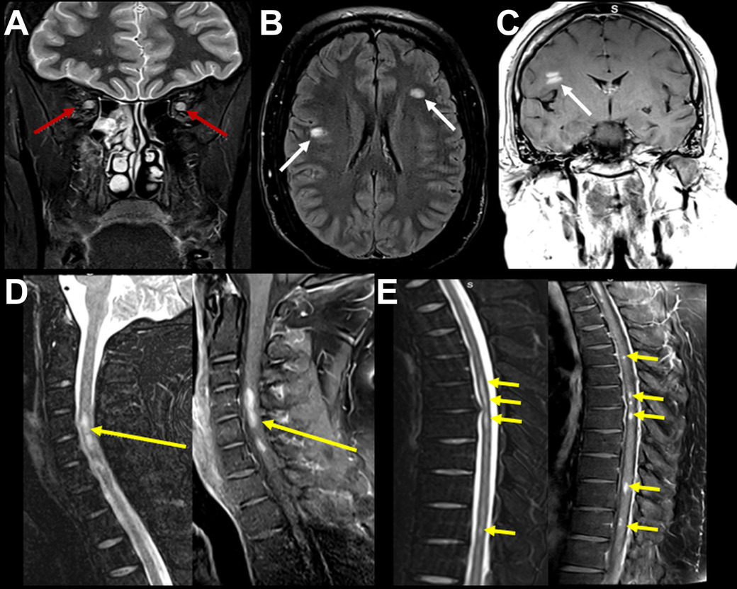
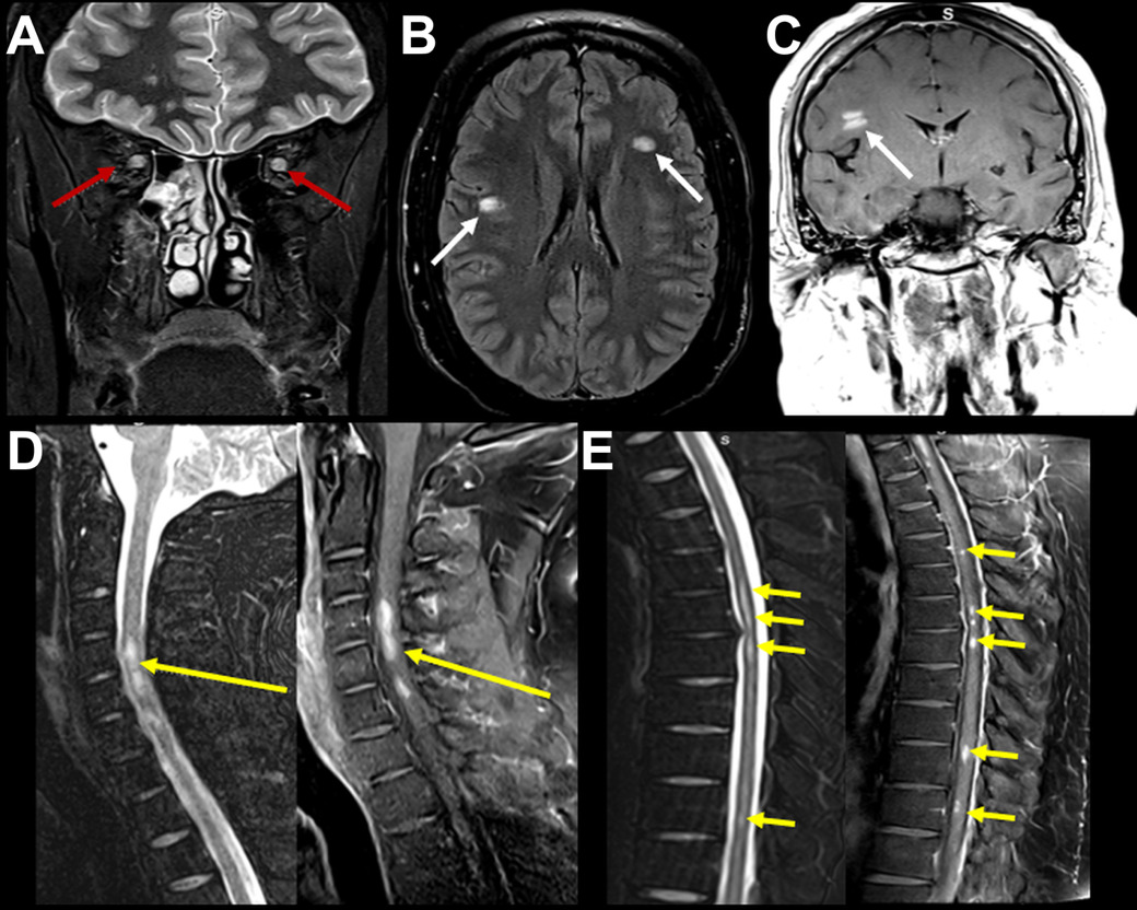
His cutaneous and hepatic GVHD were quiescent. Neurologic examination documented bilateral lower-extremity weakness and gait ataxia in association with blurred vision. Brain magnetic resonance imaging (MRI) demonstrated multifocal subcortical fluid-attenuated inversion recovery (FLAIR) hyperintensities with patchy enhancement (Figure 1). Spine MRI showed enhancing intramedullary lesions at the C4–5, T6, and T10 levels with mild cord expansion. Orbital MRI demonstrated bilateral optic nerve short tau inversion recovery (STIR) hyperintensity and enhancement, consistent with acute optic neuritis. No restricted diffusion, hemorrhage, or leptomeningeal enhancement was identified, and no imaging features of posterior reversible encephalopathy syndrome were present.
Cerebrospinal fluid (CSF) analysis revealed an elevated opening pressure of 38 cm H₂O. CSF was clear and colorless, with 4 nucleated cells/µL (5% neutrophils, 73% lymphocytes, and 22% monocytes), 5 red blood cells/µL, protein of 33 mg/dL, and glucose of 59 mg/dL. CSF cytology and flow cytometry were negative for malignant cells. Cytology and flow cytometry showed no leukemic cells. Infectious studies were negative, including CSF polymerase chain reaction for John Cunningham virus (JCV) and human herpesvirus 6 (HHV-6), as well as [cytomegalovirus, Epstein–Barr virus, herpes simplex virus 1/2, varicella-zoster virus, enterovirus, Toxoplasma gondii, cryptococcal antigen, fungal and bacterial cultures, and acid-fast bacilli. Serum [HHV-6 and JCV / Epstein–Barr virus] viral loads were undetectable. Autoimmune testing, including aquaporin-4 and myelin oligodendrocyte glycoprotein antibodies by cell-based assay in serum and CSF was negative. Bone marrow biopsy demonstrated full donor chimerism without evidence of recurrent leukemia. Brain biopsy was not pursued because the clinical presentation, multifocal imaging pattern, inflammatory CSF findings, and negative investigation for infectious, neoplastic, and alternative inflammatory etiologies collectively supported an immune-mediated demyelinating process.
The patient received intrathecal hydrocortisone, intravenous methylprednisolone 1 g daily for 5 days, and IVIG 0.4 g/kg daily for 5 doses, followed by 5 plasma-exchange sessions due to worsening lower-extremity weakness and blurred vision. Rituximab 375 mg/m2 was subsequently administered, with documented doses at approximately 1 month. Maintenance therapy consisted of monthly IVIG and oral prednisone, initially at 80 mg daily. At the 2-month follow-up, his lower-extremity weakness and blurred vision had improved substantially, allowing a reduction in prednisone to 60 mg daily. At 3 months, the patient remained clinically stable without new neurologic symptoms, and prednisone was tapered to 40 mg daily. Repeat 4-month follow-up MRI of the neural axis demonstrated near-complete resolution of the enhancement with residual T2 signal abnormality in the brain (not shown). At 5 months, vision and motor strength remained stable without recurrent optic neuritis or worsening weakness, permitting tapering to 20 mg daily. At approximately 8 months, no new neurologic episodes had occurred, and further prednisone tapering was planned. The combination of multifocal cerebral lesions, short-segment spinal cord lesions, bilateral optic neuritis, inflammatory CSF findings, antecedent multiorgan GVHD, exclusion of leukemic relapse and infection, and objective response to immunosuppression supported the diagnosis of immune-mediated demyelinating central nervous system GVHD.
Central nervous system graft-versus-host disease (CNS GVHD) is an uncommon but increasingly recognized complication of allogeneic HSCT. Reported incidences range from less than 1% in older series to 2.5% in a recent single-center cohort that applied prospective diagnostic criteria, suggesting that the entity is underdiagnosed rather than truly exceptional. Although GVHD most commonly affects the skin, liver, gastrointestinal tract, and lungs, involvement of the central nervous system has been reported in both acute and chronic forms of the disease. The diagnosis remains challenging because no single clinical, laboratory, or imaging finding is pathognomonic, and the condition frequently mimics more common post-transplant complications, including leukemic relapse, opportunistic infection, medication-related neurotoxicity, and other inflammatory demyelinating disorders.1–7
The pathophysiology of CNS GVHD is not fully understood but is thought to involve infiltration of the central nervous system by donor-derived immune cells, disruption of the blood-brain barrier, and immune-mediated injury to myelin and neural tissues. Histopathologic studies have demonstrated perivascular lymphocytic infiltrates, microglial activation, and demyelination in affected patients, supporting an inflammatory mechanism analogous to GVHD occurring in other organs. Because these processes may occur months or even years after transplantation, neurologic manifestations often arise long after initial transplant-related complications have resolved and characteristically emerge as systemic immunosuppression is withdrawn or tapered.2–4
Clinical presentations are heterogeneous and may include encephalopathy, seizures, focal neurologic deficits, optic neuritis, transverse myelitis, ataxia, and multifocal demyelinating syndromes. In a retrospective cohort of 770 allogeneic HSCT recipients, Denk and colleagues proposed three CNS-cGVHD phenotypes—vasculitis-like, demyelinating, and meningoencephalitic—and developed a 10-point scoring system that categorizes diagnostic likelihood as unlikely (1–2 points), possible (3–4 points), or probable (≥5 points). Meningoencephalitis predominated, whereas demyelinating involvement occurred in only 3 of 19 affected patients and represented the sole phenotype in only 1. Applying the demyelinating cGVHD criteria, our patient received 1 point for a compatible acute/subacute clinical syndrome, 2 points for MRI lesions involving multiple characteristic locations, 1 point for antecedent extra-CNS cGVHD, and 1 point for documented clinical improvement with immunosuppressive therapy, yielding a total score of 5 and classification as probable demyelinating CNS-cGVHD. The inflammatory CSF criterion was not met because the leukocyte count was 4 cells/µL and CSF-specific oligoclonal bands were not documented. Although neurologic symptoms developed approximately 16 months after transplantation, the relationship to cessation or reduction of immunosuppressive therapy could not be established; therefore, this criterion was not scored. Electrophysiologic and histopathologic confirmation were also unavailable. These proposed phenotypes broadly align with the cerebrovascular, demyelinating, and immune-mediated encephalitic manifestations described by the 2010 consensus conference, which emphasized the presence of extra-CNS cGVHD and exclusion of alternative etiologies.3,6,7
Neuroimaging findings are similarly variable. Brain MRI may demonstrate multifocal T2-weighted and FLAIR hyperintense white matter lesions with or without contrast enhancement, whereas spinal MRI can reveal enhancing intramedullary lesions and cord expansion. Optic nerve involvement, although uncommon, has been reported and may manifest as nerve enlargement, T2/STIR hyperintensity, and post-contrast enhancement. In the present case, the simultaneous involvement of the brain, spinal cord, and optic nerves produced a radiologic pattern resembling other inflammatory demyelinating disorders.1–4
CNS GVHD most often arises in patients with established or antecedent extra-CNS GVHD, and in the largest reported series the overwhelming majority of affected patients had concurrent or prior involvement of classic target organs; isolated CNS disease without any extra-CNS manifestation is described but distinctly uncommon and remains a diagnosis of exclusion. This distinction is practically important, because current diagnostic frameworks — including the 2010 consensus criteria and the scoring system of Denk et al. — assign explicit weight to GVHD at other NIH-defined organ sites. In our patient, the documented history of cutaneous and hepatic GVHD therefore did more than establish alloreactive susceptibility: it satisfied a formal diagnostic element, raised the pretest probability of an alloimmune CNS process over a coincidental primary demyelinating disease, and, together with the temporal relationship to immunosuppression withdrawal, materially strengthened the diagnostic assessment.2,3,6,7
The differential diagnosis is particularly important in patients undergoing HSCT. Leukemic relapse must be excluded because central nervous system recurrence may present with enhancing lesions and neurologic deficits. Opportunistic infections, including viral, fungal, and parasitic diseases, may produce multifocal imaging abnormalities in immunocompromised hosts and often require extensive microbiologic evaluation; in a patient with antecedent hemorrhagic cystitis and hypogammaglobulinemia, progressive multifocal leukoencephalopathy due to JCV and HHV-6 encephalitis warrant specific exclusion, as both may produce multifocal white matter disease and neither is reliably detected without targeted CSF polymerase chain reaction. Calcineurin inhibitor neurotoxicity and posterior reversible encephalopathy syndrome should also be considered, although the multifocal enhancing subcortical, spinal, and optic nerve lesions in our patient were incompatible with the typical posterior-predominant vasogenic edema of that syndrome. Furthermore, autoimmune demyelinating disorders such as multiple sclerosis, neuromyelitis optica spectrum disorder, and myelin oligodendrocyte glycoprotein antibody-associated disease may closely resemble CNS GVHD clinically and radiologically. In our patient, negative infectious studies, negative CSF cytology and flow cytometry, absence of recurrent leukemia on bone marrow examination, and negative aquaporin-4 and myelin oligodendrocyte glycoprotein antibodies supported an immune-mediated GVHD-related process.3–5
Because no universally accepted diagnostic criteria exist, the diagnosis of CNS GVHD is generally based on compatible neurologic symptoms, evidence of systemic GVHD or prior allogeneic transplantation, characteristic neuroimaging findings, exclusion of alternative etiologies, and response to immunosuppressive therapy. Recognition of this entity is clinically important because treatment differs substantially from that of infection or malignancy recurrence. High-dose corticosteroids remain the cornerstone of therapy, while intravenous immunoglobulin, plasma exchange, rituximab, and other immunosuppressive agents have been used in refractory cases with variable success. Most reported patients respond to immunosuppression — 14 of 19 in the cohort of Denk et al. — but response is often incomplete, and long-lasting neurologic sequelae were documented in 8 of 19, underscoring that early diagnosis and treatment may improve neurologic recovery and reduce the risk of permanent disability.4–7
This case highlights the importance of considering CNS GVHD in patients who develop new neurologic deficits months to years after allogeneic HSCT, particularly as immunosuppression is being withdrawn. Although rare, CNS GVHD should be included in the differential diagnosis when MRI demonstrates multifocal demyelinating lesions involving the brain, spinal cord, or optic nerves. Comprehensive evaluation with neuroimaging, CSF analysis including targeted testing for JCV and HHV-6, and bone marrow assessment is essential to exclude leukemic relapse and infectious etiologies before initiating immunomodulatory therapy. Early recognition of this entity may facilitate timely treatment and improve neurologic outcomes.
Disclosures/Conflicts of Interest
None
Corresponding author
Márcio L Duarte MD, Msc, PhD,
Radiology, DASA (Brazil)
Radiology, Universidade de Ribeirão Preto
Email: marcioluisduarte@gmail.com