Background
Gastric pneumatosis (GP) is defined as intramural gas in the stomach.1 The entry of air into the gastric wall can be caused by mechanical force, pulmonary disease, bacterial infection, and ischaemic condition.1 There are two conditions associated with gastric pneumatosis: Emphysematous Gastritis (EG) and Gastric Emphysema (GE). While the two conditions have similar clinical presentations, it is important to differentiate between them as both have significantly different management and prognosis.2–4 Gastric emphysema is a benign finding with an excellent prognosis, while emphysematous gastritis is a catastrophic finding with a mortality rate of more than 60%.5 Both these entities are extremely rare, and there have been very few case reports published on both. Increased use of cross-sectional imaging has led to more frequent recognition of gastric pneumatosis, including clinically benign cases of GE. Careful integration of imaging findings, clinical presentation, and risk factors may allow successful conservative management even in patients initially suspected to have EG. Despite overlapping radiographic appearances, prompt differentiation between these entities is critical to guide appropriate management and avoid unnecessary endoscopic or surgical intervention. We hence report two cases, one of emphysematous gastritis and the other of gastric emphysema, and highlight the differences in their diagnosis and management. Our cases underscore the growing recognition that not all cases of gastric pneumatosis need to be treated equally.
Cases
Case 1: Emphysematous Gastritis (EG)
An 82-year-old white male presented with a 2-day history of generalized abdominal pain, nausea, 3 episodes of vomiting, loose stools, and worsening abdominal distension. Medical history was significant for lung cancer, coronary artery disease status post coronary artery bypass grafting, heart failure with preserved ejection fraction, chronic kidney disease stage 3a, chronic obstructive lung disease, prediabetes, hiatal hernia, and erosive gastritis. There was no history of previous abdominal surgery, alcohol abuse, or nonsteroidal anti-inflammatory drug (NSAID) misuse, except for an endoscopy 3 years ago confirming erosive gastritis. The patient was on long-term proton pump inhibitors for erosive gastritis, oral pantoprazole 40 mg twice daily. Initial evaluation at home by a paramedic found him ill-appearing and diaphoretic. He was bradycardic (heart rate 42 beats per minute) and hypotensive (92/34 mm of Hg, with a mean arterial pressure of 53 mm of Hg). En route to the emergency department, he received Atropine 0.5 g and 500 mL of intravenous (IV) fluids with improvement in both heart rate and blood pressure. He also received IV ondansetron 4 mg for vomiting.
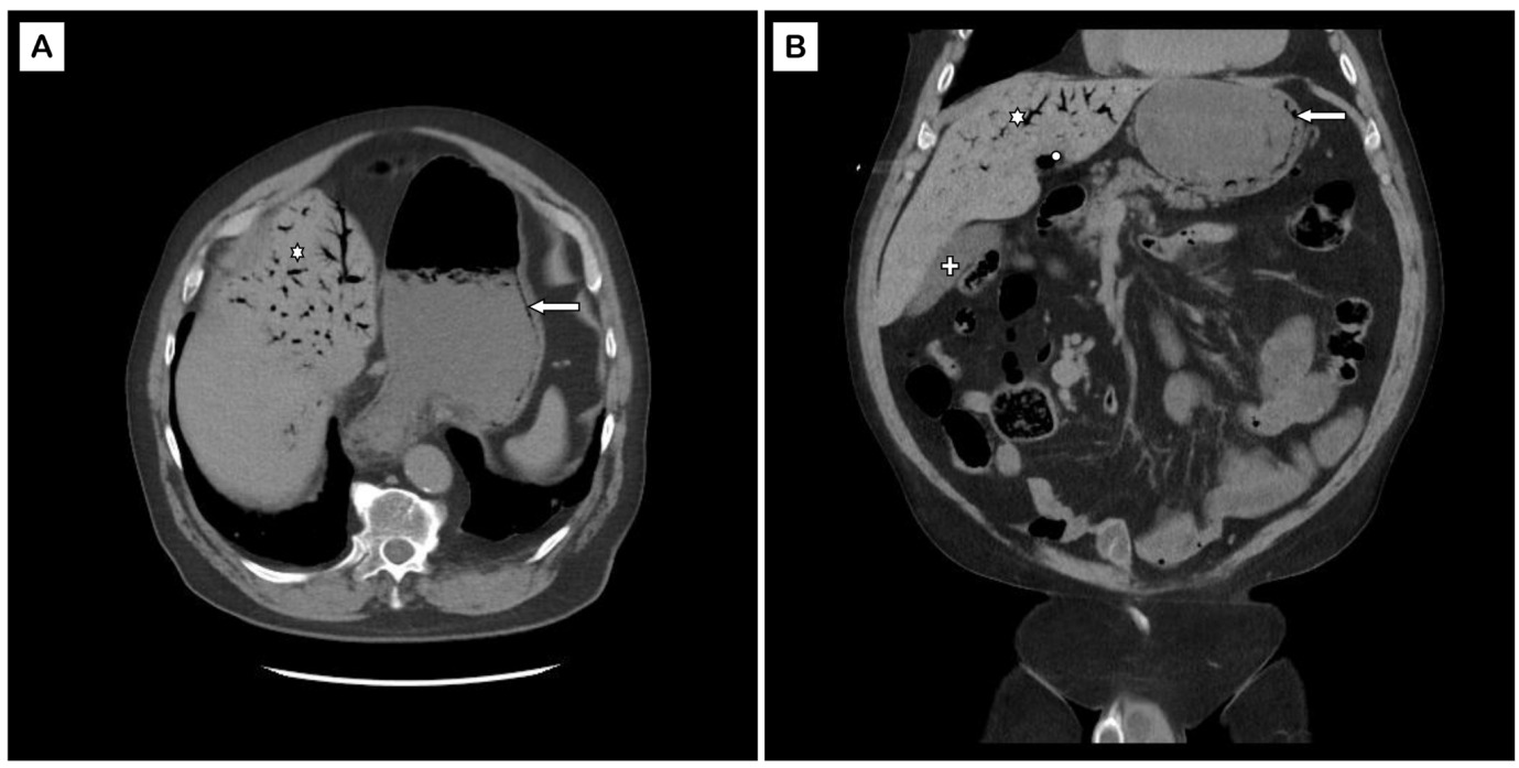
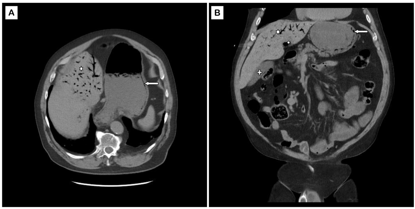
On arrival at the emergency department, his temperature was 35.9°C, heart rate 64 beats/min, blood pressure 123/59 mm of Hg, oxygen saturation of 88% on room air, requiring him to be placed on a nasal cannula at 4L/min. On examination, he was alert and awake. He had a diffusely distended abdomen with generalized tenderness. Lab tests revealed leukocytosis (14,000 cells//mm3, with 86% neutrophils), acute kidney injury (serum creatinine 1.7 mg/dL), and serum lactate 1.4 mmol/L. Blood cultures remained negative. Imaging (Figure 1) showed intramural gastric air and gastric wall thickening consistent with EG and left portal vein gas. The presence of leukocytosis, abdominal tenderness, portal venous gas, and gastric wall thickening favored a diagnosis of EG over GE despite the absence of frank sepsis. Esophagogastroduodenoscopy (EGD) was not performed due to the high risk of gastric perforation. Although blood cultures remained negative and endoscopy was deferred, the diagnosis of emphysematous gastritis remained the most likely explanation because of the combination of diffuse gastric wall thickening, portal venous gas, leukocytosis, abdominal tenderness, and hemodynamic instability at presentation. Blood cultures are frequently negative in emphysematous gastritis, and tissue confirmation is not routinely required when characteristic clinical and radiologic findings are present.
The patient was treated with IV antibiotics - ceftriaxone (later escalated to cefepime) and metronidazole. A Nasogastric (NG) tube was placed for gastric decompression. She was advised to have bowel rest by nil per os (NPO). Serial exams were performed with conservative treatment for 3 days. Given his clinical symptoms and labs improved, no intervention was deemed necessary. The NG tube was removed, and the diet was gradually advanced to regular. The patient was discharged on oral antibiotics (cefdinir and metronidazole) to complete a total duration of 2 weeks. The patient was followed in the clinic with complete clinical recovery.
Case 2: Gastric Emphysema (GE)
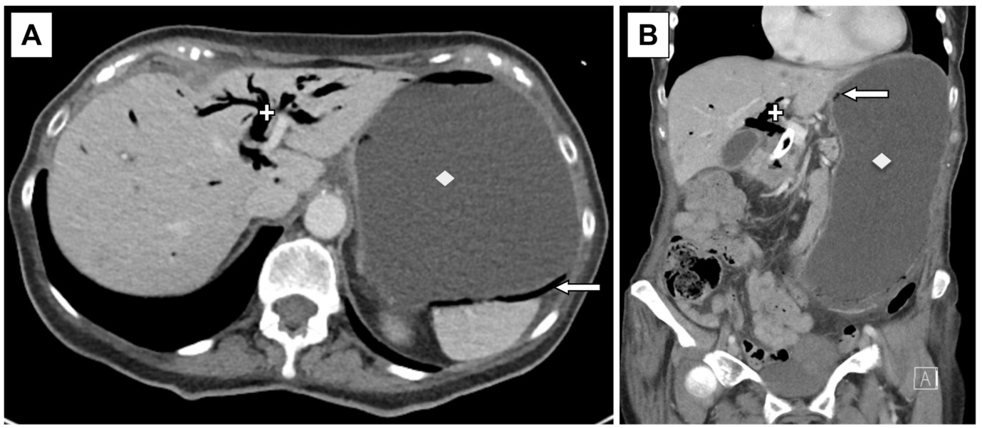
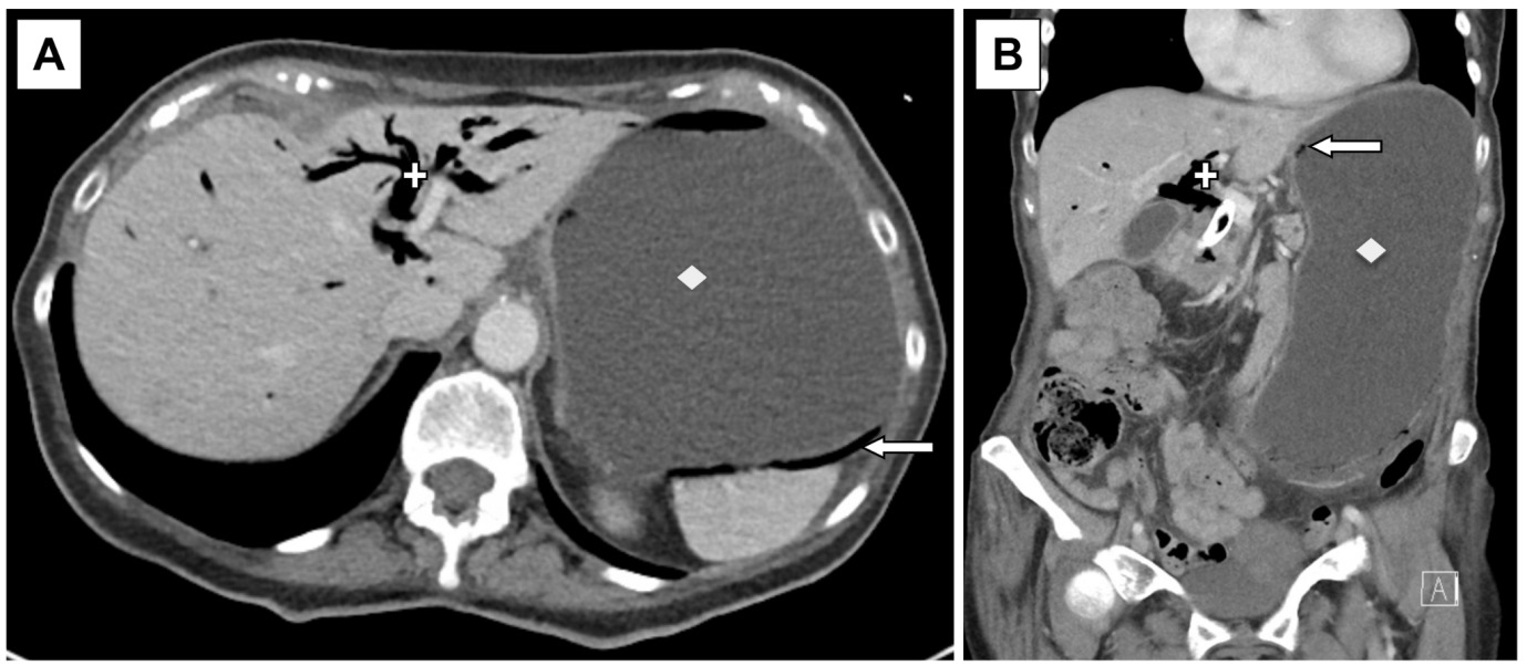
A 77-year-old white female presented with a 5-day history of worsening epigastric pain, constipation, poor oral intake, and generalized weakness. Medical history was significant for a recent diagnosis of pancreatic adenocarcinoma for which she underwent Whipple’s surgery, which was aborted due to liver metastasis, and a recent history of left lower extremity deep venous thrombosis. On arrival at the emergency department, her temperature was 36.4°C, heart rate 77 beats/min, blood pressure 155/82 mm of Hg, and oxygen saturation of 100% on room air. On examination, she was alert and awake. She had a diffusely distended abdomen but no tenderness. Laboratory workup revealed the serum leukocyte count of 12000 cells/mm3 (70% neutrophils), serum creatinine of 0.5 mg/dL, potassium 3.6 mmol/L, and lipase of 43 IU/L. Blood and urine cultures were negative. Imaging (Figure 2) of the abdomen and pelvis showed a markedly distended stomach with air within the gastric wall, suggestive of gastric pneumatosis and pneumobilia. A large necrotic mass was noted in the pancreatic head, which caused obstruction of the duodenum and distention of the common bile duct and gallbladder with pericholecystic fluid collections.
Given the clinical presentation and the absence of gastric wall thickening, hemodynamic instability, systemic toxicity, and inflammatory mucosal changes, the patient was classified as GE secondary to malignant gastric outlet obstruction. Although she was placed on empiric intravenous antibiotics (ceftriaxone and metronidazole), the antibiotics were discontinued after negative blood cultures. For bowel rest, the patient was made nil per os (NPO). A NG tube was placed for gastric decompression due to distension from gastric outlet obstruction. On day 4, she underwent EGD with endoscopic duodenal stent placement to relieve duodenal obstruction. Patient improved and was discharged on day 7 with a recommendation for a stent diet. In outpatient follow-up, she was intolerant to chemotherapy, and according to her wishes, she was eventually transitioned to palliative care for the metastatic pancreatic adenocarcinoma.
Discussion
Gastric pneumatosis is defined as the presence of gas within the stomach wall, resulting from air entry due to mechanical forces, pulmonary disease, bacterial infection, or ischemic conditions.6 Patients present with gastrointestinal symptoms, including nausea, vomiting, epigastric abdominal pain, abdominal distension, and/or tenderness, without signs of peritonitis.2 The two differentials associated with gastric pneumatosis are emphysematous gastritis and gastric emphysema, and it is challenging yet crucial to distinguish between these two conditions. The key clinical, radiologic, and management differences between EG and GE are summarized in Table 1.
Emphysematous gastritis is a life-threatening infection characterized by diffuse inflammation of the stomach wall with gas-forming organisms.7 It is associated with several immunocompromising comorbid conditions such as alcohol abuse, corrosive ingestion, recent surgery, NSAID use, dialysis, and diabetes mellitus.8 EG originates from a septic focus within the gastric wall and can be caused by various pathogens, including Klebsiella pneumoniae, Escherichia coli, Enterobacter spp., Pseudomonas aeruginosa, Candida spp., and Mucormycosis.2 While the exact pathophysiology of EG is unclear, studies suggest that the pathogenesis may include a condition such as Helicobacter pylori gastritis or a systemic immune compromise. These conditions disrupt normal barrier functions and acutely impair acid secretion, hence allowing the pathogenic organisms to colonise the otherwise sterile environment of the stomach.2 Injury to the gastric mucosal barrier resulting from corrosive ingestion, ischemia, alcohol abuse, diabetes mellitus, instrumentation, or severe inflammation permits invasion of the gastric wall by gas-producing microorganisms. Less commonly, angioinvasive fungal infections such as mucormycosis directly invade gastric tissue, causing vascular thrombosis, ischemia, and secondary gas formation. The combination of mucosal injury, impaired host immunity, and microbial invasion leads to intramural gas accumulation, inflammation, and in severe cases, transmural necrosis.2,4,5 Accordingly, patients often present with systemic signs of infection, including fever, leukocytosis, and hemodynamic instability.
Serum lactate is an important prognostic biomarker in emphysematous gastritis. Several retrospective studies have demonstrated that an initial serum lactate concentration ≥2 mmol/L is associated with significantly increased mortality and a higher likelihood of requiring surgical intervention, whereas patients with lactate levels <2 mmol/L frequently experience favorable outcomes with conservative management. In our patient, the normal serum lactate (1.4 mmol/L) provided additional reassurance that extensive transmural ischemia was unlikely and supported continued non-operative management.2,4 While imaging findings in emphysematous gastritis may closely resemble those of gastric emphysema, the presence of gas in the hepatic and portal venous systems, gastric wall thickening, and extra-gastric air in other bowel segments supports the diagnosis of EG.2,9 Recent studies have also identified the absence of marked gastric dilatation on computed tomography as an independent favorable prognostic factor, suggesting a lower likelihood of gastric ischemia and transmural necrosis.2,4
Studies suggest that all cases of gastric pneumatosis should be started initially on conservative treatment with empiric broad-spectrum antibiotics and bowel rest with NG tube placement.2 Because emphysematous gastritis is usually polymicrobial and frequently involves enteric gram-negative bacilli together with anaerobic organisms, empiric therapy should provide broad-spectrum coverage. Recommended regimens include piperacillin-tazobactam, cefepime plus metronidazole, or a carbapenem, with subsequent de-escalation according to microbiologic data and clinical response.2 Although bacterial pathogens account for most cases, fungal organisms, including Candida species and Mucorales, have been reported, particularly in severely immunocompromised patients, those with uncontrolled diabetes mellitus, prolonged neutropenia, hematologic malignancy, or persistent clinical deterioration despite adequate antibacterial therapy. Empiric antifungal therapy is therefore not routinely recommended but should be considered in these high-risk clinical settings or when fungal infection is microbiologically suspected.2,4
While EGD plays a crucial role in guiding treatment decisions, it should be performed with caution, considering clinical judgment and imaging findings to assess the risk of perforation. Although a rare occurrence, patients with EG may not always require surgical intervention and can recover with non-surgical management, as seen in our case. Historically, emphysematous gastritis carried mortality rates exceeding 50–60%, leading many patients to undergo urgent surgery. However, accumulating evidence supports that patients without perforation, diffuse peritonitis, uncontrolled sepsis, or extensive gastric necrosis may be successfully managed with aggressive conservative therapy consisting of bowel rest, gastric decompression, intravenous fluids, proton-pump inhibition, and broad-spectrum antimicrobial therapy together with close clinical monitoring.5 Surgical intervention should be considered for patients who develop gastric perforation, transmural necrosis, uncontrolled hemorrhage, diffuse peritonitis, abdominal compartment syndrome, or persistent clinical deterioration despite maximal medical therapy.10 Operative procedures generally involve partial or total gastrectomy with resection of nonviable tissue, occasionally requiring damage-control surgery in critically ill patients. Although postoperative mortality remains substantial, carefully selected patients undergoing timely surgery for irreversible gastric necrosis may achieve improved survival.
Gastric emphysema, on the other hand, is a rare benign condition occurring in patients due to several predisposing conditions that can be classified as either obstructive, pulmonary, or traumatic. Obstruction distal to the stomach causes gastric distention and mucosal tears, allowing the air to diffuse into the gastric wall, leading to gastric emphysema. Some of the causes of obstruction include peptic ulcer disease, carcinoma, duodenal obstruction secondary to various conditions, pyloric stenosis, and gastric volvulus.3,11 Pulmonary pathologies such as asthma, chronic obstructive pulmonary disease, pneumothorax, etc., lead to increased intrapulmonary pressure. This leads to gastric emphysema due to alveolar rupture and air penetrating the gastric wall from the mediastinum.3,11–13 Lastly, iatrogenic traumatic injuries during endoscopies, gastrostomies, and NG tube placement are the most common cause of gastric emphysema.11 Therefore, a thorough history and careful examination are essential to identify these underlying etiologies. Other risk factors for gastric emphysema include ischemia, atherosclerosis, hypertension, alcoholism, and diabetes mellitus.14 The imaging typically demonstrates linear or circumferential intramural gastric gas without significant gastric wall thickening. Although imaging may reveal findings such as portal venous gas and pneumatosis intestinalis,13 as seen in our case, where imaging showed pneumobilia, EGD remains the definitive diagnostic tool, confirming the absence of inflammatory mucosal changes. These patients are typically managed conservatively with bowel rest, gastric decompression, IV fluids, and treatment of the underlying cause. They do not require surgery unless there are necrotic changes in the mucosa.
In conclusion, gastric pneumatosis represents a rare but clinically significant radiologic finding encompassing two distinct entities with markedly different prognoses. This finding includes both benign and life-threatening etiologies. Portal venous gas and gastric wall thickening increase concern for severe gastric injury, like emphysematous gastritis. But, it should always be interpreted within the overall clinical context and in conjunction with accompanying gastric wall imaging findings, laboratory abnormalities, and systemic manifestations, as it may also occur in benign gastric emphysema. Absence of systemic toxicity is likely linked to gastric emphysema. Although emphysematous gastritis is traditionally associated with high mortality, early recognition and prompt conservative management may result in favorable outcomes in selected patients. Conversely, gastric emphysema is generally benign and often resolves with treatment of the underlying mechanical cause. Awareness of the distinguishing clinical and radiographic features of these entities is essential to optimize management and avoid unnecessary surgical intervention.
Acknowledgements
We would like to thank our librarian, Erika Schulz-Vendrella, for providing academic support.
Disclosures/Conflicts of Interest
None
Corresponding author
Sushrut Ingawale, MD, DNB, MBBS
Internal Medicine Physician,
Department of Medicine,
Brigham and Women’s Hospital and Harvard Medical School,
Boston, Massachusetts, United States
Email: singawale@bwh.harvard.edu