A 41-year-old male with a history of exploratory laparotomy following a gunshot wound 20 years ago presented to the emergency department with abdominal pain and vomiting of 1-week duration. He reported progressively worsening postprandial epigastric pain associated with daily episodes of non-bilious, non-bloody vomiting occurring immediately after meals. The review of symptoms was otherwise negative. On examination, the patient appeared malnourished and emaciated. The physical examination revealed a midline scar with no evidence of abdominal tenderness, mass, or distension. Serum creatinine was elevated at 2.02 mg/dL (normal 0.6–1.3 mg/dL). Hemoglobin was elevated at 18.1 mg/dl, consistent with hemoconcentration. Other laboratory investigations did not reveal any significant abnormality. The patient had been hospitalized one year earlier with a similar presentation of epigastric pain and vomiting. At that time, CT imaging raised concern for intussusception or a possible obstructing mass at the duodenal bulb. The upper endoscopy demonstrated a large inflammatory lesion at the junction of the first and second portions of the duodenum. Histopathologic evaluation of endoscopic biopsies revealed duodenal mucosa with chronic inflammation and prominent Brunner’s glands consistent with Brunner gland hyperplasia (BGH), without evidence of dysplasia or malignancy. The patient improved with conservative management and was discharged with proton pump inhibitor therapy but was subsequently lost to follow-up.
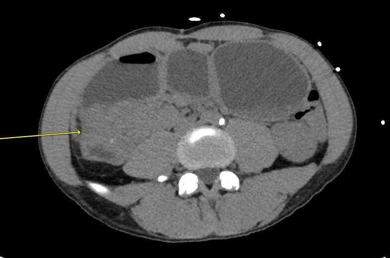
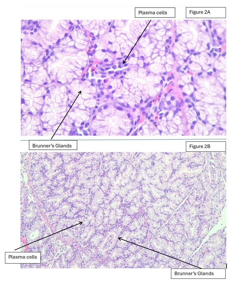
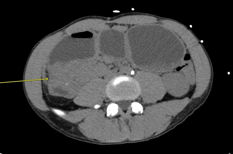
A CT scan performed during this admission revealed an ill-defined soft tissue mass measuring 5.5 x 4.2 cm at the gastric pylorus and proximal duodenum, with loss of fat plane between duodenum and pancreas, causing GOO, as shown in Figure 1. Multiple enlarged lymph nodes were also noted, highly suspicious for a neoplastic etiology. As the patient was lost to follow-up, we were unable to determine whether lymphadenopathy resolved. Upper GI endoscopy during the current admission revealed a hypertrophic duodenal mass amenable to biopsy. As shown in Figure 2, histopathology results from the duodenal mass again revealed a benign Brunner’s gland nodule but with superficial erosion and mild active duodenitis, while the stomach biopsy revealed gastric mucosa with mild inactive chronic gastritis, intestinal metaplasia with no dysplastic changes or evidence of H. pylori infection.
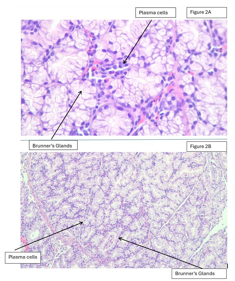
Histologic examination demonstrated a nodular and lobular proliferation of closely packed Brunner glands involving the deep mucosa and submucosa, with intervening fibromuscular septa. The glands were composed of bland mucinous epithelial cells with small, basally located nuclei and no significant cytologic atypia or mitotic activity. No dysplasia or invasive carcinoma was identified. Focal surface erosion and mild active duodenitis were present. Immunohistochemical staining showed a low Ki-67 proliferative index and a wild-type p53 staining pattern. The patient was initially managed with nasogastric tube decompression and slowly advanced to a liquid diet. He was discharged to follow up with a hepatobiliary surgeon for the resection of the hypertrophic duodenal mass lesion and possible gastrojejunostomy. The patient lost follow-up on discharge.
BGH is a rare, benign condition of the duodenum, first described in 1835, with fewer than 200 cases reported in the literature.1 Brunner’s glands are predominantly located in the proximal duodenum and play a protective role by secreting alkaline mucin to neutralize gastric acid. BGH is most often identified incidentally during endoscopic or radiologic evaluation performed for unrelated indications.2 While the majority of cases are asymptomatic, BGH can become clinically significant depending on lesion size, morphology, and anatomical location. Symptomatic patients may present with nausea, vomiting, dyspepsia, epigastric pain, upper gastrointestinal bleeding, or, rarely, gastric outlet obstruction.3,4 Brunner’s glands, located in the submucosal layer of the duodenum, secrete alkaline mucin to protect the duodenum from stomach acid.5 Hyperplasia of these glands is believed to result from increased inflammation or excessive stomach acid production and is associated with conditions such as H. pylori infection, chronic pancreatitis, and uremia.6 The incidence of BGH decreases as one move further from the duodenal bulb.7
Most cases of BGH are asymptomatic and detected incidentally during endoscopic or radiologic evaluation. Common symptoms include dyspepsia, vomiting, epigastric pain, and, rarely, GOO or gastrointestinal bleeding.3,5,7,8The size, location, and type of BGH determine these features.8 In our case, epigastric pain, vomiting, and poor oral intake were attributed to GOO on CT imaging. The differential diagnosis includes both benign and malignant conditions, such as duodenal adenocarcinoma, pancreatic head carcinoma, gastrointestinal stromal tumors, neuroendocrine tumors, lymphoma, and inflammatory lesions. Distinguishing these entities based solely on imaging findings can be difficult, particularly when features such as wall thickening, mass effect, or lymphadenopathy are present. Consequently, tissue diagnosis remains essential when evaluating suspicious duodenal lesions.6,9
Diagnosis of BGH is challenging and ultimately requires histopathologic confirmation.10Endoscopically, BGH may appear as a nodular, polypoid, or thickened duodenal mass, which can mimic malignancy.1Although CT may show a duodenal mass distinct from the pancreas, it often cannot reliably distinguish BGH from malignancy, as seen in our case.6
Tissue histopathology remains the definitive method for diagnosing BGH.9,11Because Brunner gland proliferations predominantly involve the submucosa, superficial endoscopic biopsies may be nondiagnostic or may underestimate the extent of the lesion.9,11 Endoscopic ultrasound (EUS) may provide valuable additional information by defining the layer of origin and the relationship of the lesion to adjacent structures, particularly the pancreatic head.4,11 When routine endoscopic biopsies are inconclusive, EUS-guided tissue acquisition may improve diagnostic characterization.4 These modalities may help avoid unnecessary radical surgery in patients with benign Brunner gland lesions.4,11,12 Routine forceps biopsies may be nondiagnostic because Brunner gland proliferative lesions frequently involve deep mucosa and submucosa and may be covered by intact or nonspecific surface mucosa.9,11EUS can define the lesion’s layer of origin, depth of involvement, and relationship to the pancreatic head and may help distinguish an intrinsic duodenal lesion from an adjacent pancreatic process.4,11 EUS-guided fine-needle aspiration has been reported to establish the diagnosis of Brunner gland hyperplasia in lesions initially suspected to represent pancreatic malignancy.4 Therefore, when forceps biopsy findings are nondiagnostic or discordant with imaging, EUS-guided fine-needle aspiration/biopsy or another deep-tissue sampling technique may provide more representative tissue.4,11 Given rare reports of dysplastic or malignant change arising in association with Brunner gland lesions.13,14 limited biopsy specimens may not completely exclude an unsampled neoplastic component in a large or radiologically progressive lesion. Misdiagnosis of BGH has been reported. In some cases, this has led to unnecessary aggressive treatment, including pancreatoduodenectomy (Whipple procedure). These decisions were often based on misleading biopsy or imaging findings. Preoperative biopsies may yield variable results, ranging from normal tissue to adenocarcinoma. Concerning CT findings, they have also contributed to misdiagnosis. These include loss of the fat plane between the duodenum and pancreas and lymph node enlargement.6,8,12
Despite two benign endoscopic biopsy results, definitive intervention was considered because of recurrent mechanical obstruction, inability to maintain adequate oral intake, clinical evidence of malnutrition, progressive radiologic abnormalities, and persistent inability to exclude an unsampled malignant process. The feasibility of endoscopic resection was also limited by location. The management of Brunner gland lesions depends on symptoms, lesion size and location, feasibility of endoscopic resection, and the degree of concern for malignancy.10,11 Symptomatic lesions causing obstruction or gastrointestinal bleeding generally warrant definitive removal, with endoscopic resection considered when technically feasible.5,10,11 Surgical management is generally reserved for lesions that cannot be safely or completely resected endoscopically or for cases in which malignancy remains a significant concern despite diagnostic evaluation.3,10,12
Prior case reports have described BGH presenting as duodenal or pancreaticoduodenal-region masses that mimic malignancy.1,3,6,12 In the present case, progressive radiologic evolution over time, including increasing mass size, loss of the fat plane between the duodenum and pancreas, and associated lymphadenopathy, further heightened concern for malignancy and increased diagnostic uncertainty. Additionally, recurrence of symptoms after prior conservative management increased suspicion for neoplastic disease. The majority of BGH cases (approximately 99%) are benign. However, some papers have reported its possible hypothetical evolution into dysplasia.13 Rare malignant transformation or coexisting malignancy cannot be completely excluded, particularly given the limitations of biopsy and the aggressive radiologic appearance.2,3,6,12,14 EUS was not performed, and the available medical record did not document a specific reason for its omission.
BGH, although rare, should be considered in the differential diagnosis of duodenal masses presenting with GOO. Imaging findings may closely mimic malignant processes, particularly when associated with lymphadenopathy or loss of fat planes. This case highlights the limitations of radiologic assessment alone and underscores the importance of multimodal evaluation, including endoscopic ultrasound and adequate tissue sampling, to establish an accurate diagnosis. Recognition of this entity is essential to prevent unnecessary radical surgical procedures.
Disclosures/Conflicts of Interest
None
Corresponding Author
Hansika Sharma, MD
Internal Medicine Resident
WellStar Spalding Medical Center
601 8th Street, Griffin, Georgia, USA, 30224
Email ID: hansikas0209@gmail.com