A 47-year-old woman with stage IV estrogen receptor positive, progesterone receptor positive, and human epidermal growth factor receptor 2 positive (ER+/PR+/HER2+) breast cancer with known liver metastases, presented to the emergency department with abdominal pain. The liver metastases were first diagnosed 24 months prior. She reported several weeks of worsening abdominal distention and pain, persistent nausea with intermittent non-bloody emesis, and recent unintentional weight loss with early satiety and fatigue. She endorsed mild constipation due to chronic opioid use. She had no prior liver or gallbladder disease, alcohol use, or known hepatotoxic exposures. Family history was notable for Breast Cancer (BRCA) gene negative breast cancer in her mother. Her oncologic treatment consisted of docetaxel, trastuzumab, and pertuzumab followed by endocrine therapy, with subsequent courses of trastuzumab, deruxtecan, docetaxel, fulvestrant, and abemaciclib for progressive hepatic metastases.
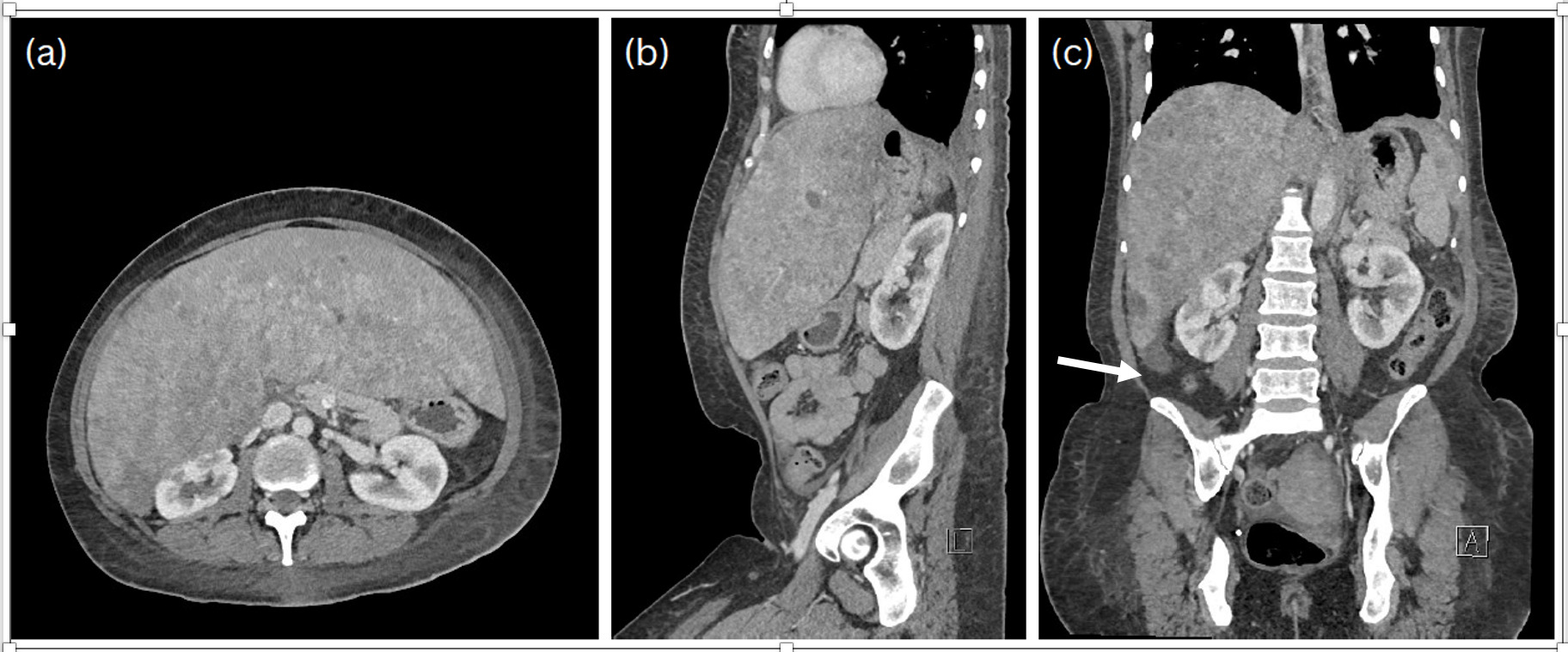
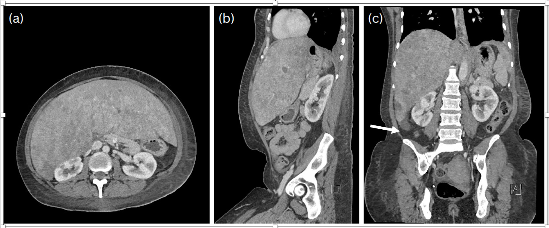
On physical exam, she appeared cachectic. Her abdomen was distended with right upper quadrant tenderness and palpable hepatomegaly. Contrast-enhanced computed tomography (CT) of the abdomen and pelvis demonstrated marked liver enlargement compared to prior imaging, with a span measuring at least 26 centimeters and extending caudally below the iliac crests with innumerable metastatic lesions replacing most of the hepatic parenchyma (Figure 1 a, b, c). Laboratory evaluations over the prior 9 months demonstrated elevation of Alkaline phosphatase (ALP) and preserved hepatic synthetic function. ALP ranged from 132-338 [reference range: 13-35], aspartate aminotransferase (AST) 12-53 U/L [13-35], alanine aminotransferase (ALT) 12-53 U/L [7-45], total bilirubin 0.4-0.6 mg/dL [0.2-1.0], albumin 3.7-4.3 g/dL [3.2-4.8], prothrombin time (PT) 12.3-15.0 sec [12-13.8] and international normalized ratio (INR) 0.92-1.18 [0.8-1.2].
Despite extensive hepatic metastatic disease on imaging, her laboratory findings were discordant. Mostly reflecting milder physiological and pathological changes and relatively preserved hepatic synthetic function. We suggest that this was due to several factors. The liver demonstrates substantial functional reserve and regenerative capacity, maintaining metabolic and synthetic function even with up to 80% parenchymal replacement by tumor provided that remaining hepatocytes remain viable.1 Tumor metastases form expansive nodules and displace rather than eradicate hepatocytes. Remaining viable cells facilitate compensatory hypertrophy of non-malignant hepatic parenchyma and increase metabolic activity per cell.1–3 As tumor burden increases, the liver actively compensates for space-occupying disease. The adjustment of liver size to 100% of what is required for homeostasis has been called “hepatostat”. This mechanism occurs exclusively in the liver and can maintain metabolic capacity even in advanced metastatic disease.2,3 Hepatic progenitor cells, cytokines, growth factors and other mechanisms are involved in this unique process. Acute liver regeneration occurs after loss of hepatocytes. Additional regenerative pathways become activated when normal regeneration is impaired and trigger the appearance of “progenitor” cells. Chronic loss of hepatocytes is associated with regenerative efforts characterized by continual hepatocyte proliferation and over time often results in cirrhosis and/or liver cancer.3 Additionally, liver transaminases also reflect hepatocellular injury or cholestasis rather than synthetic or functional capacity, limiting their ability to accurately represent overall disease burden.4 Similarly, ALP increases with biliary obstruction or canalicular injury, which may not reflect the extent of parenchymal replacement.5 ALP elevation may also occur due to bone metastases, systemic inflammation and prolonged immobility which further reduces its specificity in this setting. Her preserved serum albumin level, despite cachexia supports the above mechanisms. It is probable that her younger age, lack of alcohol use, adequate baseline nutritional status and other comorbidity also protected her from more severe liver dysfunction.
Our case suggests that preserved hepatic function can occur despite severe tumor burden. Imaging findings, clinical symptoms, and trends in laboratory studies should be interpreted together when evaluating patients with advanced metastatic malignancy. This can guide clinicians’ decision-making, prognostication, and discussions of goals of care. We also suggest maintaining a high index of suspicion for hepatic decompensation in patients with progressive metastatic disease, even when standard liver function tests initially appear relatively preserved.
Disclosures/Conflicts of Interest
None
Corresponding author
Michael E. Lazarus, MD
Professor of Clinical Medicine
757 Westwood Plaza, Suite 7501C, Los Angeles, CA 90095
Email: MLazarus@mednet.ucla.edu