Background
Opportunistic pulmonary infections cause significant morbidity and mortality after solid organ transplantation (SOT), as chronic immunosuppression impairs host defenses and broadens the range of pulmonary pathogens.1–3 In liver transplant recipients, persistent infiltrates require a broad differential diagnosis, including bacterial pneumonia, fungal infection, cytomegalovirus, Mycobacterium tuberculosis, nontuberculous mycobacteria (NTM), Nocardia, and malignancy.2,3 Tuberculosis (TB), nontuberculous mycobacteria (NTM), and Nocardia each present diagnostic and therapeutic challenges in immunocompromised hosts. Latent tuberculosis reactivation may go undetected because screening tests have reduced sensitivity in this population, while rapidly growing NTM and Nocardia can mimic malignancy with nodular or cavitary lesions, requiring targeted microbiologic testing and prolonged therapy. Rifamycin-tacrolimus interactions further complicate management and require close trough monitoring. We report concurrent pulmonary detection of M. tuberculosis, M. fortuitum, and N. beijingensis in a liver transplant recipient, complicated by hyponatremia from syndrome of inappropriate antidiuretic hormone secretion (SIADH) and significant drug interactions.4–12
Case Presentation
A 66-year-old Chinese man with chronic hepatitis B treated with tenofovir disoproxil fumarate 300 mg orally daily, status post liver transplantation approximately 17 months before this presentation, hypertension, and atrial fibrillation presented with progressive functional decline after a prolonged hospitalization one month earlier for multifocal pneumonia after outpatient cefdinir failed. His home immunosuppressive regimen consisted of tacrolimus 4 mg every morning and 3 mg every evening, with no other anti-rejection agents. During that initial admission, he received intravenous piperacillin-tazobactam every eight hours for seven days and underwent bronchoscopy, which demonstrated no endobronchial lesions or mucosal abnormalities. The infectious workup included negative Streptococcus pneumoniae and Legionella urinary antigens, a negative respiratory viral panel, negative serum beta-D-glucan, negative serum galactomannan, and negative serum antibody testing for human immunodeficiency virus type 1 (HIV-1) and type 2 (HIV-2). The QuantiFERON result was indeterminate; multiple sputum evaluations for M. tuberculosis and the bronchoalveolar lavage (BAL) M. tuberculosis polymerase chain reaction (PCR) were negative. Routine respiratory and BAL cultures were initially reported as negative; these were interim results while mycobacterial cultures remained in prolonged incubation. The patient was discharged without antibiotics. After discharge, his functional status worsened, and he required a walker, whereas he had previously been independent in activities of daily living.
On readmission, initial laboratory evaluation revealed profound hyponatremia with serum sodium 105 mmol/L (reference range, 135-145 mmol/L), and he was admitted to the critical care unit for treatment with 3% sodium chloride. The patient was clinically euvolemic. Laboratory findings met standard diagnostic criteria for SIADH, including hypotonic hyponatremia with serum osmolality 247 mOsm/kg (reference range, 281-297 mOsm/kg; diagnostic threshold <275 mOsm/kg), inappropriately concentrated urine with urine osmolality 236 mOsm/kg (reference range, 40-1400 mOsm/kg; diagnostic threshold >100 mOsm/kg), and natriuresis with random urine sodium 37 mmol/L (no reference range applicable for a random specimen; diagnostic threshold >30 mmol/L). Serum uric acid of 2.7 mg/dL (reference range, 3.5-7.2 mg/dL; supportive threshold <4 mg/dL) further supported the diagnosis. Severe hypothyroidism, a secondary cause of euvolemic hypotonic hyponatremia, was excluded with a normal thyroid-stimulating hormone of 0.89 uIU/mL (reference range, 0.55-4.78 uIU/mL). His sodium level improved, and he was transitioned to oral sodium chloride tablets before transfer from the critical care unit to telemetry. A sputum culture obtained during the prior hospitalization identified Mycobacterium fortuitum. Susceptibility testing for M. fortuitum showed susceptibility to amikacin, ciprofloxacin, doxycycline, moxifloxacin, and trimethoprim-sulfamethoxazole; intermediate susceptibility to cefoxitin and linezolid; and resistance to clarithromycin and tobramycin.
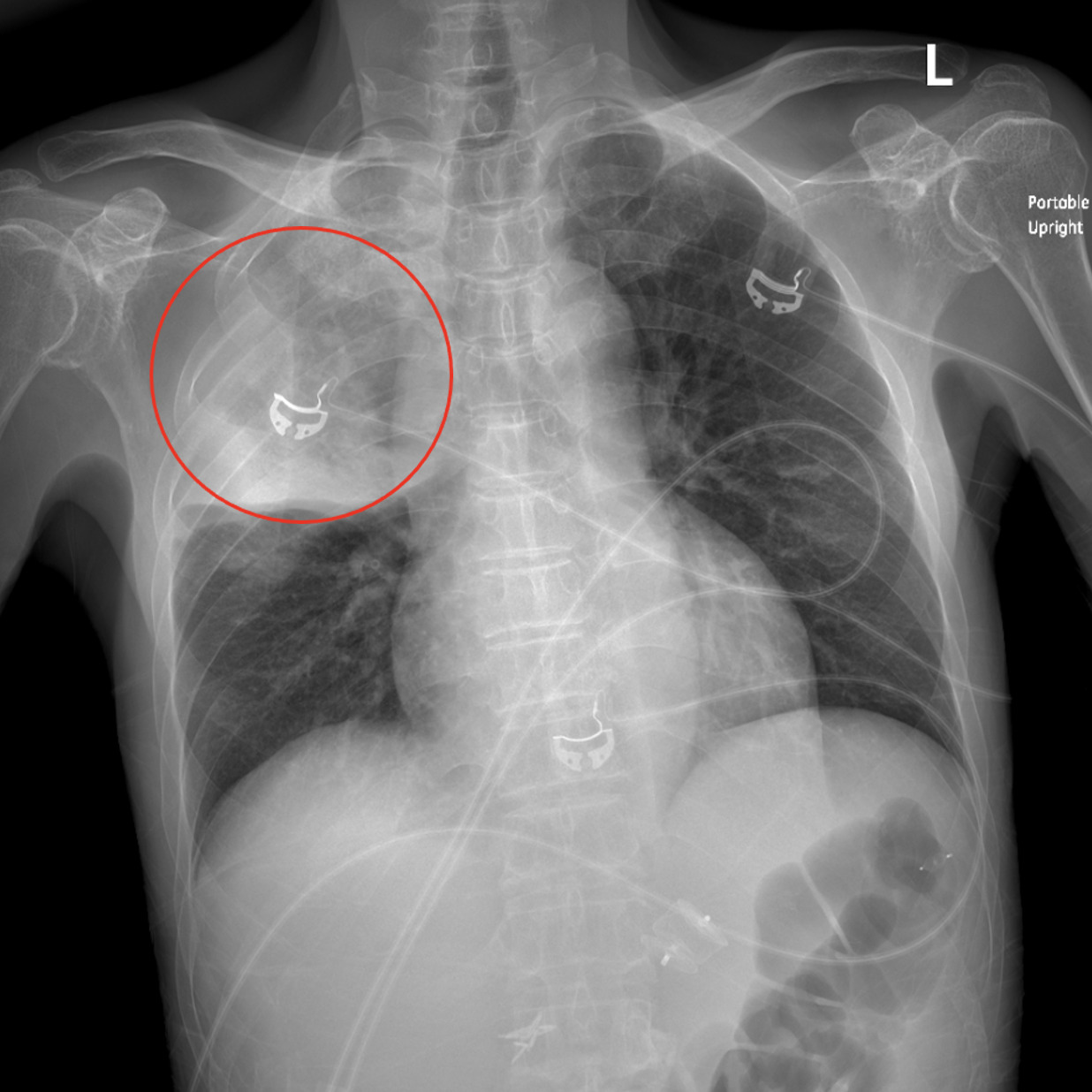
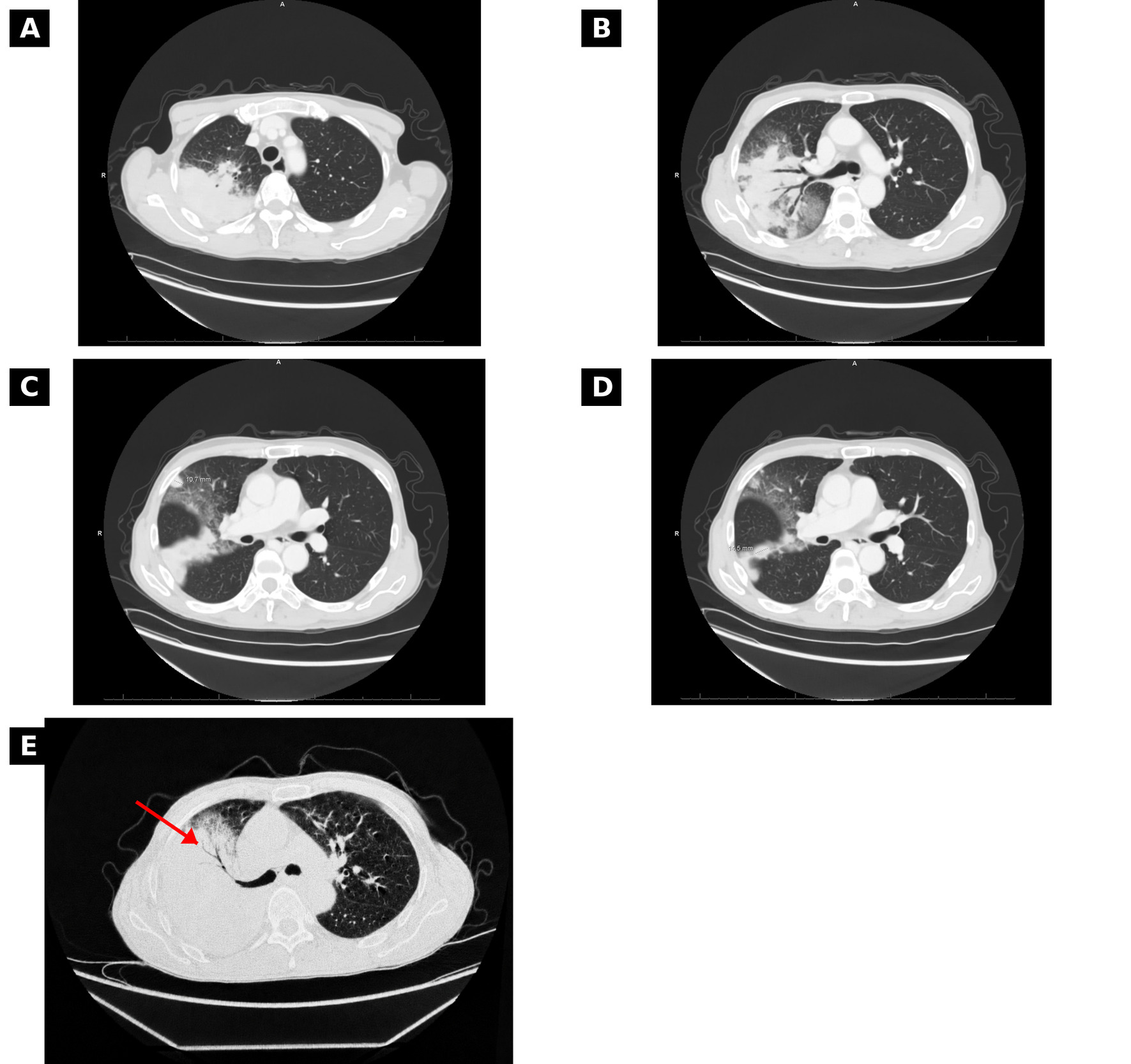
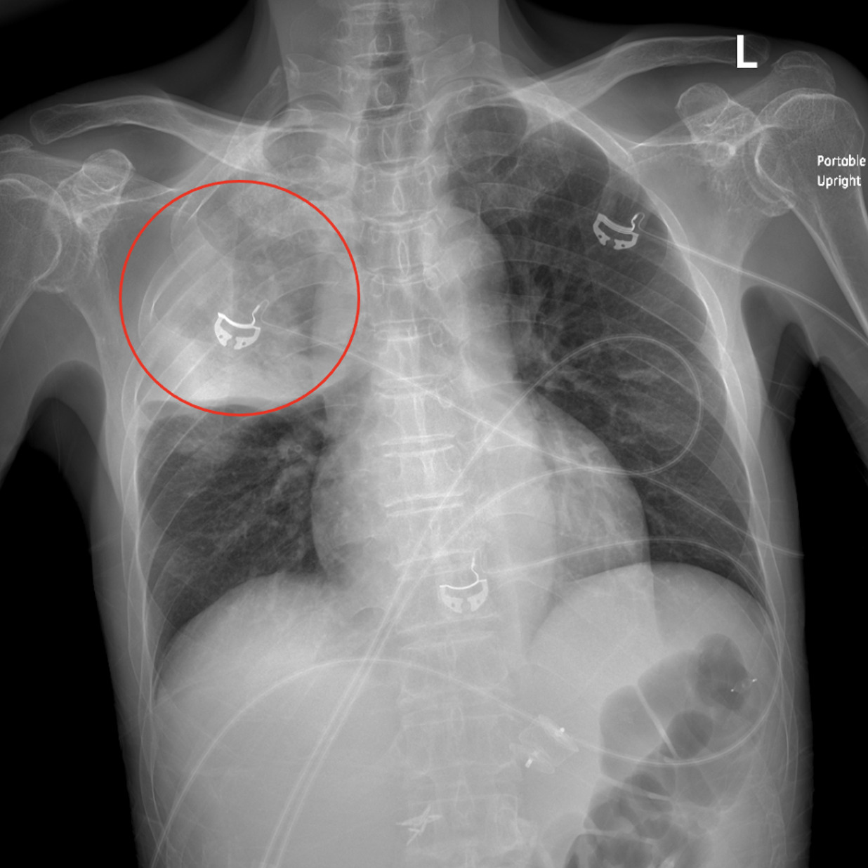
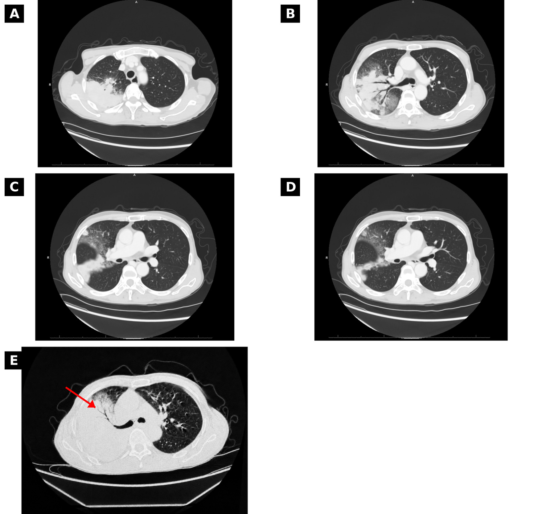
Chest radiography obtained on readmission showed dense right upper lobe air-space consolidation with volume loss (Figure 1). The contrast-enhanced chest computed tomography (CT) from the previous admission showed large areas of right upper lobe consolidation with alveolar and ground-glass opacities and possible small masses measuring up to 1.5 cm along the major fissure. A non-contrast chest CT obtained on readmission showed progression to near-complete right upper lobe consolidation with air bronchograms, concerning for advanced infection with possible underlying malignancy (Figure 2). Brain CT obtained the following day showed bilateral low-density subdural collections favored to represent hygromas or chronic subdural hematomas, without acute intracranial hemorrhage. Neurosurgery did not recommend any acute intervention.
Several days into the readmission, because the right lower lobe nodule was considered the most technically feasible and accessible target for percutaneous biopsy, interventional radiology sampled that lesion. Frozen section showed inflammation without malignancy. Final pathology showed inflammatory changes without malignancy or caseating granulomas. Shortly after the biopsy, an acid-fast bacillus (AFB) culture from BAL obtained during the prior hospitalization completed its prolonged incubation and identified Mycobacterium tuberculosis complex. This delayed growth occurred despite the indeterminate QuantiFERON result, negative BAL M. tuberculosis PCR, and multiple negative sputum evaluations for M. tuberculosis during the prior admission. Tissue culture from the lung biopsy remained in incubation and was later identified as Nocardia beijingensis. Susceptibility testing showed susceptibility to amikacin, ceftriaxone, clarithromycin, doxycycline, imipenem, and trimethoprim-sulfamethoxazole, and resistance to amoxicillin/clavulanate and ciprofloxacin.
In the setting of immunosuppression and progressive pulmonary disease, distinguishing active infection from colonization or contamination was difficult. M. tuberculosis recovered from a pulmonary specimen was considered clinically significant and unlikely to represent contamination. Although M. fortuitum and Nocardia can represent colonization in respiratory samples, the patient’s immunocompromised state, radiographic progression, and recovery of N. beijingensis from lung tissue led Infectious Disease consultants to treat both as true pathogens. Shortly after M. tuberculosis was identified, the patient was transitioned from empiric piperacillin-tazobactam to intravenous imipenem and oral trimethoprim-sulfamethoxazole, moxifloxacin, isoniazid with pyridoxine supplementation, pyrazinamide, and rifabutin. Rifamycin-mediated induction of tacrolimus metabolism required close dose adjustment and trough monitoring. His home tacrolimus regimen was 4 mg every morning and 3 mg every evening, and at discharge it was 5 mg twice daily. He was transferred to a tertiary care center for further management by his transplant team.
Discussion
This case highlights the complexity of evaluating non-resolving pulmonary infiltrates in a solid organ transplant recipient. The initial routine cultures were negative at the time of reporting, but subsequent finalization of mycobacterial cultures and tissue culture demonstrated concurrent detection of M. tuberculosis, M. fortuitum, and N. beijingensis. M. tuberculosis was considered clinically significant, and recovery of N. beijingensis from lung tissue supported invasive infection. M. fortuitum and N. beijingensis were treated as pathogens because of the host’s immunosuppression, radiographic progression, and tissue recovery of N. beijingensis; however, the available conventional microbiologic data could not definitively establish the relative contribution of each organism or exclude colonization by one isolate. This sequence illustrates the importance of following delayed microbiologic results and pursuing tissue diagnosis when radiographic progression raises concern for malignancy or atypical infection.7–10,13–15 Concurrent recovery of M. tuberculosis, M. fortuitum, and N. beijingensis is rare but biologically plausible during chronic post-transplant immunosuppression. These organisms may produce overlapping nodular, cavitary, or mass-like pulmonary findings.
The initial regimen was selected to provide activity against TB, rapidly growing NTM, and Nocardia while accounting for transplant-specific toxicity and drug interactions. It included imipenem, trimethoprim-sulfamethoxazole, moxifloxacin, isoniazid with pyridoxine, pyrazinamide, and initially rifabutin. A later switch to rifapentine was recalled but could not be confirmed. Treatment required monitoring for hepatotoxicity, myelosuppression, renal dysfunction, electrolyte abnormalities, neurologic toxicity, and antimicrobial-immunosuppressant interactions.6–9,11,12 The rifamycin-tacrolimus interaction was a central management issue. Rifampin and rifapentine are potent cytochrome P450 3A4 (CYP3A4) inducers and can markedly reduce tacrolimus exposure, risking subtherapeutic immunosuppression and graft rejection unless tacrolimus doses are adjusted with frequent trough monitoring.6,11,12 The patient required stepwise tacrolimus dose adjustments in response to low trough levels, illustrating the need for coordinated care among Infectious Diseases, Gastroenterology/Transplant Hepatology, and the transplant team. This case reinforces the importance of balancing infection control with graft preservation in liver transplant recipients receiving multidrug antimicrobial therapy.
This report has several limitations. The available records did not confirm pre-transplant interferon-gamma release assay (IGRA) or tuberculin skin testing, or prior latent-TB treatment; the treating physician noted that screening would have been routine but did not have access to the transplant records. The patient did not report trimethoprim-sulfamethoxazole prophylaxis on the admission medication reconciliation or in external prescription records, but earlier post-transplant use at Jackson could not be excluded. Conventional cultures established organism recovery but could not quantify each isolate’s relative contribution or fully distinguish active infection from colonization. After transfer, only the tertiary hospital admission note was available. Attempts to obtain follow-up were unsuccessful, precluding verification of the final antimicrobial regimen, treatment durations, clinical response, and long-term outcome.
In this case, integrating delayed respiratory culture results with lung-biopsy cultures prevented premature closure of the diagnostic evaluation. Management required continued diagnostic assessment, attention to delayed culture finalization, biopsy of an accessible pulmonary lesion, broad antimicrobial therapy, and careful management of rifamycin–tacrolimus interactions. In solid organ transplant recipients with persistent pulmonary infiltrates, clinicians should maintain a broad differential, obtain targeted testing for mycobacteria and Nocardia when appropriate, and interpret multiple positive cultures in the context of the clinical presentation and specimen source.
Acknowledgments
The authors thank the patient and the multidisciplinary care team, including Infectious Disease, Gastroenterology/Transplant Hepatology, Pulmonology, Interventional Radiology, and Neurosurgery, for their contributions to the patient’s care.
Disclosures / Conflicts of Interest
The authors declare they have no conflicts of interest. The authors acknowledge the use of an artificial intelligence (AI)-based writing assistant to support reference formatting and manuscript organization, in accordance with journal policy. The authors are responsible for all clinical content, interpretation, and final wording.
Corresponding Author
Anthony Membreno
Herbert Wertheim College of Medicine, Florida International University
11200 SW 8th Street, AHC2, Miami, FL 33199, USA
Tel: (305) 348-0570
Email: amemb005@fiu.edu